All posts by Nafis Hasan

Notes from the North – MMCRI Transgenic Core

MMCRI transgenic core

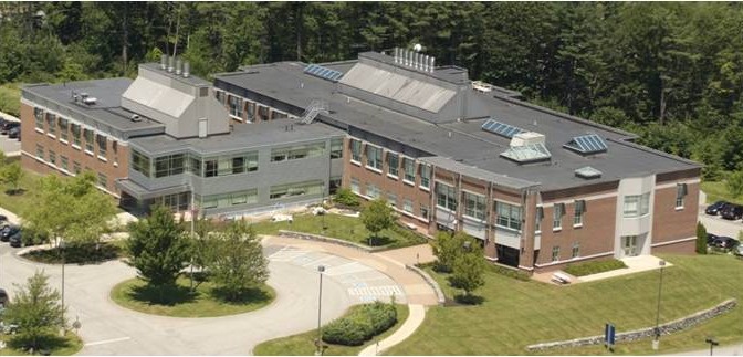
The Maine Medical Center Research Institute (MMCRI) has partnered with Tufts to provide a professional core facility that has over 15 years of experience providing high quality services for the generation of mouse transgenic strains including the use of CRISPR/Cas, cryopreservation of mouse germ cells, and imaging, including MRI and microCT. Mice are generated in a full barrier, AAALAC-accredited animal facility in a transgenic production room that facilitates direct importation of mice into the Tufts barrier facility. Contact us to discuss your mouse and imaging projects.
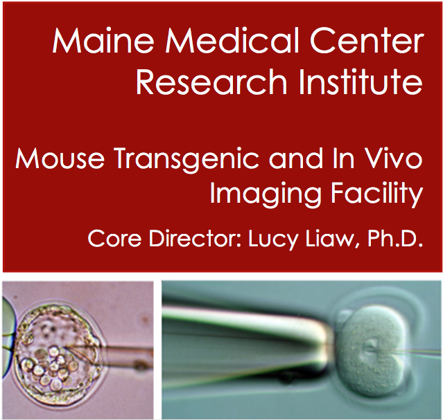
Mouse Transgenesis

Source - MMCRI transgenic core
Source – MMCRI transgenic core

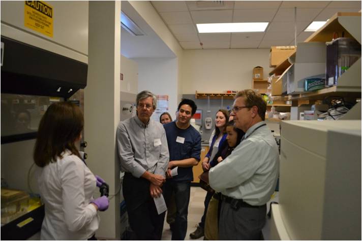
We provide microinjection to generate your mouse models. Services include microinjection of fertilized oocytes with traditional DNA transgenes, or microinjection of CRISPR/Cas. ES cell injection is also performed. Contact us to design your CRISPR mouse project – cost depends on type of modification, strain of mouse, and days of injection. We have a high surveillance production room that will allow importation of mice direct into some barrier facilities.

Contact – Lucy Liaw, Ph.D., liawl@mmc.org
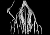
microCT

Source - MMCRI transgenic core
Source – MMCRI transgenic core

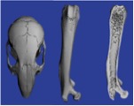
We house a Scanco high speed in vivo microCT scanner X-ray system. Our microCT facility has extensive experience in bone imaging and quantification, and can work on other projects where tissues are provided, i.e., vascular imaging of samples perfused with microfil. We provide quantification and any 3D images of the samples as required. Contact us to get a project quote. Pricing is based on hours of scanning and analysis time.

Contact – Lucy Liaw, Ph.D. liawl@mmc.org
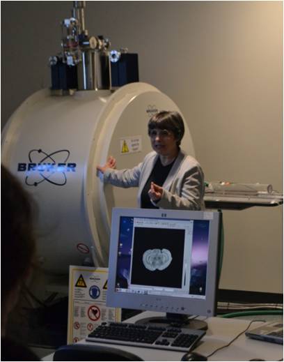
Small Animal MRI

Source - MMCRI transgenic core
Source – MMCRI transgenic core

Our MRI facility houses a Bruker Pharmascan 7T, 300 MHz imager with 100 μm resolution. Services include anatomical imaging of most organs, angiography, proton spectroscopy and localized spectroscopy, and cardiac imaging, including diastolic and systolic dimensions of the ventricle. We can house “clean” animals at our facility for studies requiring longitudinal imaging. Contact us for mor information.

Contact – Ilka Pinz, Ph.D. pinzi@mmc.org

Nobel Laureate Dr. Susumu Tonegawa to speak at HNRCA

Susumu Tonegawa
Susumu Tonegawa, Ph.D. (Source – tonegawalab.org)


The Jean Mayer Human Nutrition Research Center (HNRCA) on Aging has invited the Nobel laureate Dr. Susumu Tonegawa to give a talk as part of the Drs. Joan and Peter Cohn Family lecture on nutrition, inflammation and chronic disease. This event also includes a panel discussion besides presentations from other distinguished speakers such as Jonathan Kipnis, Ph.D. (Director, Center for Brain Immunology and Glia, University of Virginia), Simin Meydani (Director, HNRCA), Irwin Rosenberg, M.D. (Director, Nutrition and Neurorecognition lab, HNRCA) and Dennis Steindler, Ph.D. (Director, Neuroscience and Aging lab, HNRCA). This event will take place on Dec 17, 2015 from 1-6 pm at the HNRCA building. (Registration link – https://secure.www.alumniconnections.com/olc/pub/TUF2/events/event_order.cgi?tmpl=events&event=2363222)


Dr. Susumu Tonegawa is currently the director of RIKEN-MIT Center for Neural Circuit Genetics and holds the Picower professorship of Biology and Neuroscience. He was awarded the Nobel Prize in Physiology or Medicine in 1987 for his discovery of the genetic mechanism behind antibody diversity in the adaptive immune system. His experiments, that started in 1976, countered the contemporary dominant idea that each gene produced one protein as he went on to show that genetic rearrangements in mature B cells in adult mice, compared to embryonic mice, are responsible for the diversity observed in antibodies.

Dr. Tonegawa received his Ph.D. in Biology from University of California, San Diego (UCSD) in 1968. He then went on to work as a postdoctoral scholar at the Salk Insitute in San Diego, and later at the Basel Institute for Immunology in Switzerland where he performed his landmark experiments. His current research focuses on understanding the molecular, cellular and neural circuit mechanisms underlying learning and memory formation using genetically engineered mouse models that have spatially or temporally restricted neurotransmitter receptor and enzyme expression, or have conditional knock-out of specific cell populations that are suspected to be involved in memory formation. These mutant mice, along with control mice, are then subjected to analytical methods such as behavioral tasks, in vitro electrophysiology, and both in vivo and in vitro high resolution optical imaging. The main questions his research sees to answer include what happens in the brain during memory formation, consolidation of short-term memory to long-term, and memory recall. He also seeks to understand the role of memory in decision-making, and how other factors such as reward, punishment, attention and emotional state can affect learning and memory formation. These research questions have great implications in understanding memory disorders such as Alzheimer’s where patients are unable to form new memories, or PTSD where patients are unable to suppress recalling of a highly unpleasant memory.


For more details on his work, please visit http://tonegawalab.org/research/.

Caroline Genco, PhD, awarded Arthur E. Spiller, M.D. Professorship

Caroline Genco, Ph.D. (Source – Sackler website)


On Tuesday, Dec 1, 2015, Dr. Caroline Genco, chair of the Integrative Physiology & Pathobiology, was installed as the inaugural Arthur E. Spiller, M.D. Professor  This professorship was made possible by an estate gift from Dr. Spiller and this fund is meant to support “an outstanding biomedical researcher and educator at Tufts University School of Medicine who demonstrates expertise in the field of genetics”.


Dr. Genco completed her Bachelor’s degree in Biology at State University of New York (SUNY-Fredonia) and did her graduate work in microbiology at University of Rochester School of Dentistry & Medicine. She went on to work as a postdoctoral scholar at Center of Disease Control (CDC) and later, walked the path of an academic that led her to Boston University School of Medicine (BUMC) prior to Tufts. At BUMC, she was honored with the Lifetime Achievement Award for Research & Service (2012). She has also served on numerous NIH study sections, worked with & advised several pharmaceutical companies, and has mentored a host of graduate students and postdoctoral scholars throughout her career.


Dr. Genco’s research spans basic, translational and global health in relation to mucosal pathogens, with a particular interest in genetic elements of host-pathogen interactions in systemic inflammatory disease states. Some of her notable works include establishing the connection between oral mucosal bacteria and their role in atherosclerosis and plaque formation. Her current work focuses on innate immune responses to mucosal pathogens, regulatory mechanisms in bacterial pathogens and the association between the microbiome and chronic inflammation, with an interest in pancreatic cancer. More details are available at her Sackler webpage – http://sackler.tufts.edu/Faculty-and-Research/Faculty-Research-Pages/Caroline-Genco.


Besides her academic accomplishments, Dr. Genco also serves as the President and Treasurer of the Christina Clarke Genco Foundation, Inc. This non-profit was established in honor of Christina Genco, who passed away in a tragic biking accident in 2011. This organization embodies the values of late Ms. Genco and seeks to empower young adults so they can make a difference in their communities. In addition, the organization also focuses on improving biking safety, providing scholarship to female athletes and assisting affordable housing initiatives.

BIOBUGS – Call for volunteers

Are you interested in getting some teaching and outreach experience? If so, consider volunteering to participate in the fall semester’s BIOBUGS (Biology Inquiry and Outreach with Boston University Graduate Students) program. BIOBUGS is a week-long program where we invite Boston area high school biology classes to come to Boston University and participate in a newly redesigned 3 hour lab, Comparative Vertebrate Anatomy!

In this lab students will study the internal anatomy of six classes of vertebrates (cartilaginous and bony fish, amphibian, reptile, bird, and mammal) through hands-on examination of dissected whole specimens as well as prepared skeletons and skins. They will then be asked to formulate hypotheses about the ecology of each dissected species based on their observations of the similarities and differences between each specimen.
We are looking for Sackler graduate students who want to either teach or volunteer for one or more days of the program. No prior experience in vertebrate anatomy is required – instruction will be provided! The labs will be run December 10-11 and December 14-16, 2015 from 9am-12pm, and are followed by a free pizza lunch from 12pm-1pm for all teachers and volunteers. Please contact Melissa LaBonty (melissa.labonty@tufts.edu) or Joslyn Mills (Joslyn.mills@tufts.edu) to sign up or get more information.


biobugs
Credit – Frankie Velazquez, Immunology

7th Annual Tufts Neuroscience Symposium & William Shucart Lecture, 2015

Guest Post by Michaela Tolman

On Thursday October 8th, the Neuroscience Department hosted its 7th annual Neuroscience Symposium and William Shucart Lecture. The daylong event brings together neuroscience enthusiasts from the entire Tufts community, including the Departments of Neurosurgery, Psychiatry and Neurology as well as the basic science departments of Tufts University School of Medicine. The day is filled with talks celebrating the cutting edge of neuroscience research and stimulating conversations. The final lecture of the day honors William Shucart, MD. With nearly 200 people in attendance, the 2015 Symposium was a great success.

This year, Dr. Thomas Biederer of the Neuroscience department served as symposium director. Invited speakers included Dr. QiuFu Ma from Dana-Farber/Harvard Medical School: “Spinal circuits transmitting mechanical pain”, Dr Scott Soderling from Duke University Medical Center: “Actin badly – cytoskeletal drivers of neuropsychiatric disorder”, Dr. Elly Nedivi from MIT: “Structural dynamics of inhibitory synapses”, Dr. Pavel Osten from Cold Spring Harbor Laboratories: “Automated analysis of functional and anatomical circuits in the mouse brain”, and Dr. Christina Alberini from New York University: “Molecular mechanisms of memory consolidation and enhancement”. Students and post docs had the opportunity to meet the speakers in small groups during lunch and talk more in depth about their research and experiences.

The day concluded with Dr. Gordon Fishell from New York University giving the 2015 William Shucart Lecture on his work in the field of interneuron development. Dr. Philip Haydon, chair of the Neuroscience Department, describes in his welcome Dr. Shucart’s contribution to the Neuroscience Department, “…William Shucart, MD, who at that time was chair of Neurosurgery, recognized the importance of basic Neuroscience and was unwavering in his support for the formation of our Department. It is only fitting therefore that the last lecture recognizes his important contributions.” Previous Shucart Lecturers include Dr. Martha Constantine-Paton, Dr. Karl Deisseroth, and Dr. Mark Schnitzer to name a few. More information can be found on the Symposium website at http://medicine.tufts.edu/Education/Academic-Departments/Basic-Science-Departments/Neuroscience/Neuroscience-Symposium-and-Shucart-Lecture and the Neuroscience Department’s Facebook page.

Attendees at the 7th annual Neuroscience Symposium, 2015


Michaela Tolman is a Neuroscience PhD Candidate in the Yang lab, studying astrocyte maturation and functional development. She is also the current President of the Sackler Graduate Student Council. 

Sackler Science Open Mic Night

Guest Post by Alex Jones

Of the myriad skills a scientist must have in his repertoire, arguably the most important is the ability to clearly present his findings.  Whether speaking amongst colleagues, giving a talk at a scientific meeting or simply answering the age-old question, “So, what do you do?” at a cocktail party, the need for better scientific communication skills is ever present.  But how does one improve their public speaking abilities insofar as they relate to science?  The answer is as simple as it is nerve-wracking (at least for some); that is, speaking publicly about science.

With this in mind, on October 20th, the Sackler Graduate Student Council Career Paths committee and the Tufts Biomedical Business Club teamed up to present the first “Sackler Science Open Mic Night”.  The goal of the event was for students to present short, two to three minute talks covering some aspect of their research and to help each other workshop these talks, in the hopes of improving.  This “flash talk” style of presentation is challenging as it leaves only enough time for the speaker to present the most crucial aspects of their research, but it is also one of the most frequently used skills whether it be at a networking event, an interview, or even in response to that question at a cocktail party.

Professor Dan Jay joined students for the event and to kick it off he gave a flash talk of his own on a favorite subject of his, “the intersection between art and science”.  Student presenters from all over Sackler gave talks ranging from astrocytes (and their communication with neurons via vesicular release of transmitters) to v-ATPases (and the signaling pathways that control their assembly).  Presenters and spectators alike made the event a success, providing tons of feedback on how to improve those talks for future presentations.  Keep an eye out for more events similar to the Sackler Science Open Mic Night in the future as the Sackler Graduate Student Council and Tufts Biomedical Business Club look for more ways to promote scientific communication.

Dr. Dan Jay at the inaugural Science Open Mic Night

Alex Jones is a Neuroscience PhD student in the Reijmers lab studying changes in molecular profile of neurons during memory formation. He also serves as the current Treasurer of the Sackler Graduate Student Council.


Notes from the North – MMCRI Open House

Guest Post by Jessica Davis-Knowlton

About two hours north of where you sit reading your InSight there is another site of Tufts scientific discovery waiting to greet you! Tufts Sackler and Tufts University Medical School have partnerships with institutes in Maine allowing students to experience research and medicine in a unique setting with a strong emphasis on collaboration. During the 3rd and 4th rotations, first year students in Sackler have the opportunity to rotate with faculty members at the

MMCRI aerial view
MMCRI aerial view

Maine Medical Center Research Institute (MMCRI) in Scarborough, Maine before joining the CMDB program or with faculty at Jackson Laboratories (Jax) in Bar Harbor, Maine before joining the Genetics program. For TUSM students in the Maine Track Program immersion in the particular challenges facing Maine physicians starts with brief stints to Maine in the first and second years followed by a 9-month Longitudinal Integrated Clerkship in the third year.

Enough background though; what is particularly exciting about Maine this November is that MMCRI will be holding their annual Open House on the 20th and all are encouraged visit! The Open

MMCRI open house 2014

House is a great opportunity to investigate our cores (Transgenics and Gene Targeting, Histopathology and Antibody Production, Confocal Microscopy, Small Animal Imaging, Protein and Nucleic

MMCRI small animal imaging

Acid Analysis, Molecular Phenotyping, Physiology and Behavior, and Clinical and Translational Research Services/Tissue Bank), see posters from Tufts and UMaine MMCRI grad students, meet the faculty, tour the building, and of course see green mice.

For first year CMDB students coming to visit on the 20th I’d like to highlight the newest principal investigator to join MMCRI and Jumbo alum, Michaela Regan, PhD. Dr. Reagan has a B.S. in Engineering from Harvey Mudd College in Claremont, California and a Ph.D. in Biomedical Engineering from Tufts University in Medford, Massachusetts. During her graduate

Michaela Regan
Dr. Michaela Regan

research, Dr. Regan studied Breast Cancer Bone Metastasis by investigating mesenchymal stem cell (MSC) tumor homing and developed silk scaffold implants with therapeutic bone marrow MSCs that deliver anti-tumor proteins to breast tumors. Her post-doctoral fellowship was in the lab of Dr. Irene Ghobrial at the Dana-Farber Cancer Institute/Harvard Medical School where she focused on understanding how multiple myeloma cells manipulate their bone marrow niche to support their growth and cause osteolytic lesion formation. She developed a 3D model of inhibited osteogenesis in silk scaffolds and examined the roles of abnormally expressed microRNAs in MSCs in this process. She also developed bone-targeted, bortezomib-loaded nanoparticles to modulate the bone microenvironment and make it less receptive to cancer cell colonization. When you see Dr. Regan, ask her about her path to becoming a PI: it’s all about collaboration and networking folks!

MMCRI Lucy Liaw
Dr. Lucy Liaw & Collaborators

Collaboration is the name of the game up here in Maine, as research institutes scattered about the state are relatively small. As a fortuitous consequence, a culture of cooperativeness and the drive to reach far outside normal comfort zones to seek said cooperation has prevented research in Maine from becoming insular. As pressure by publishers for completeness and complexity in manuscripts mounts, partnerships between labs, and the skills to develop such partnerships, have become ever more indispensible.

Remember, Boston had more snow than Portland last year so don’t fear the “winter, still winter, almost winter, and road construction” description of seasons in Maine: come see the Maine-Tufts partnership in action!

Jessica Davis-Knowlton is a 3rd year CMDB student in the Liaw lab in MMCRI.  Her thesis work focuses on the role of Notch signaling in smooth muscle cells involved in atherosclerosis. 


Ten Cardinal Rules For Being a Lab Scientist

Guest Post by Ania Wronski, PhD.

  1. Write everything down

Whether you are learning a technique from someone or you are running you thousandth PCR reaction, if you do something different or noteworthy – write it down. I assure you, when months down the line you need to repeat a similar reaction (and you will!) you will NOT remember the conditions that you thought were so obvious that you didn’t write it down. Even details that may not seem very significant – like, I had a longer lunch so my blot was in blocking solution for 2 hours instead of one, may make a big different to the end result and knowing why may vastly improve your reproduction of the results.

  1. Be organized

When you start a project, identify a storage structure and STICK TO IT. This sounds relatively simple, but it isn’t! Are you going to organize your scientific literature by topic? What if it covers multiple topics? What filename will you use to save it? How are you going to organize your data? By experimental type? Date? Project? You want to be able to find things relatively easily and NOT have multiple copies of the same or similar files floating around.

Same goes for labelling tubes. Please, for the love of the PCR fairy, please DO NOT label your tubes 1-8. At least put the date and some redeeming feature. One of the best things to do is to log it in a database with its details and location so you have the capacity to search for it electronically – but it does take time to establish these systems and it is often MUCH harder to go back and re-organize your samples once you have 4 boxes of plasmids and several boxes of primers…

  1. Be consistent

Science is already hard – why make life harder for yourself? When you get in stock solutions, make them to a nice, repeatable number. Dilute primers to 50 or 100uM every time you get a new primer tube. That way, you don’t need to look up what you did. It is a standard dilution for all your primers. This also leads to better science as consistency is often the best way to reduce variability and increase the reproduction of results. Passage cells a certain way? Why deviate? You could be introducing variability that may influence your downstream applications.

  1. Re write protocols for a dummies guide.

You could be a cloning god or goddess, but what happens when you don’t have to do a technique for an extended period of time? You forget. Re-write your protocols so they explain all the steps, including the little quirks that you discovered are the keys to success. This is especially important for logins to shared equipment that you may not access very frequently (I.e. the nanodrop password on level 5 is DropitLikeItsHot). I often write several versions of protocols: a lengthy “extended” protocol with explanations and detailed information and a short “mini” protocol that I can just fill out the blanks as I need for the experiment. One warning about having spreadsheet protocols: you can accidentally introduce errors and not even realize it! So it’s always good to check your spreadsheet or have some “control” calculations that will let you know if something is wrong.

  1. Make sure you use the appropriate controls!

If an experiment fails, controls can help you determine if it was a technical failure or a negative result (same with the inverse – a positive result or a technical glitch). Many may think that controls are a waste of time and reagents, however controls are more important than your experimental sample because it tells you if your result can be trusted. You are only as good as your positive and negative controls!

  1. Understand your techniques

Nowadays there is a kit for everything technique conceivable – the age of convenience has hit the world of scientific research. Although kits can make your experiments much easier, they are also a very easy way to become complacent. If you don’t know how an experiment actually works, you won’t know the limitations of your results and the conclusions you can or cannot draw from them. In addition, troubleshooting is MUCH easier if you understand the rationale behind how an experiment works.

  1. Never stop learning

The vast majority of scientists has an innate desire for knowledge – it’s why we are scientists to begin with! You can learn something from everybody, from the new grad student to the seasoned postdoc. This includes the lab down the hall working on a random organism that doesn’t seem related. Some of the greatest discoveries in science came about when multiple fields crossed paths. Never turn down an opportunity to learn, as your knowledge is your best asset as a scientist.

  1. Ask for helpEveryone is always busy so it can seem daunting to ask someone with a full plate for help. However, you are surrounded by incredibly skilled and intelligent individuals. If you do not ask, you cannot ever receive. When you do ask, try to be mindful of their time, maybe even email them to ask for a good time to sit down and chat with them. Know what you want to ask and make sure it’s not an answer that can be relatively easily answered by google or a lab protocol that is easily accessed. Much of the knowledge of science is passed down from lab member to lab member.
  1. Don’t think about the how, think about the WHY
    When you are the bench, it is very easy to fall into a pattern of simply doing experiments because that feels comfortable. A great scientist will consider why they are doing an experiment. Before you rush into anything new, sit down and consider all of your data. What is it telling you? What would the next logical step be? What question are you really asking? What is the big picture? It is pointless to mindless conduct experiments if they will not help further your question and the eventual result. Even if you know it is highly unlikely that you will achieve your end goal, you should still one in mind.
  1. Be prepared for change.
    Two years ago, CRISPR sounded like a type of cracker, nowadays it’s the hot new technique. Science is dynamic and is constantly changing. You need to be prepared for these and be amendable to changes, whether it be new techniques, a change in direction or even location. If you are working in industry, it is common for projects to suddenly be terminated or drastically change, even if everything is going well. In academia, we are not as efficient at culling projects and tend to beat the proverbial dead research horse because we really want a project to work out. One critical skill of a researcher is to know when they have hit a dead end and to move on. It is extremely difficult and you often need to convince powerful people (i.e. your PI) that it’s the right decision.

Originally published on AdageOfAnia.com

 Ania is a post-doctoral fellow in the Kupperwasser lab and blogs regularly about “Science!” at www.adageofania.com

Techniques – Wild, Wild West(erns)

Need some help to tame that wild western blot? Here are some tips and tricks to help you along the way –

  • Always check ladder migration pattern based on specific gel and electrophoresis conditions, as these factors can shift the apparent molecular weights from the supposed “standard” ladder image given out by the company.
  • When testing a new antibody, leave the blot intact, opting to strip it and perform a control protein blot after probing for your target protein. Cutting the blot and using different pieces for your target and control protein on a first try may obscure alternative target protein isoforms or off-target background staining.
  • High background? Try a more stringent blocking condition than just BSA or milk by adding goat serum or fish gelatin to your solution. Blocking overnight also can help clean up your blots.
  • Is you gel “smiling” or “frowning”? This usually happens when your sample buffer has too much salt.
  • Make sure your PVDF membrane is pre-activated with methanol for 20 minutes before making gel sandwich. It’s also a good idea to mark which side of the membrane is facing the gel with a sharpie, on a top corner.
  • It’s always a good idea to do a Ponceau stain on your membrane after transfer to make sure your transfer went all right. Alternatively, you can also stain your gel with Coomassie blue.
  • It is possible to over-transfer, especially for low MW proteins (<10 kDa) – optimize transfer time or reduce voltage. On the other hand, high MW proteins will take longer time to transfer.
  • Have a dirty secondary? Consider adding a wee-bit of Tween-20 to your washes (0.01-0.5%).
  • When loading your samples, press on the pipet just enough to get any possible air bubbles out and run the tip through the running buffer in the tank before putting it in the well. And make sure your sample was denatured prior to running.
  • If power supply reading shows 0 when switched on, make sure your power cables are properly connected to the power supply. If that doesn’t work, check for broken electrodes or blown fuses. Lastly, try with a higher limit power supply.
  • As always, make sure you write down the protocol before-hand and check through every step when performing. This will help you track your steps back to see at which step things could have gone wrong.


Source - http://prooffreaderplus.blogspot.com/2014/05/one-of-my-favourite-memories-good-bad.html
Source – http://prooffreaderplus.blogspot.com/2014/05/one-of-my-favourite-memories-good-bad.html