Identification of Epitope Regions in the Ara h 2 Allergen
In “Identification and Mutational Analysis of the Immunodominant IgE Binding Epitopes of the Major Peanut Allergen Ara h 2”25, scientists analyzed the amino acid structure of Ara h 2 to determine in which regions it interacted with IgE antibodies – the locations of its epitopes.
They began by purifying Ara h 2 and electrophoresing it onto a gel, from which an amino acid sequence was obtained. From that sequence, nineteen overlapping peptides were sequenced. These peptides, all together, represented the entirely of Ara h 2, which each one fifteen residues in length and overlapping with the next by eight amino acids. A diagram showing the general idea of overlapping peptides appears in Figure 24, below. The peptides were then probed with pooled blood serum from fifteen peanut-allergic patients (as well as a control patient with no peanut allergy). In the regions where binding epitopes were present, a positive response was recorded by the blood serum probe. In Figure 24, binding regions are indicated by yellow stars and the hypothetical probe results are shown to the right.
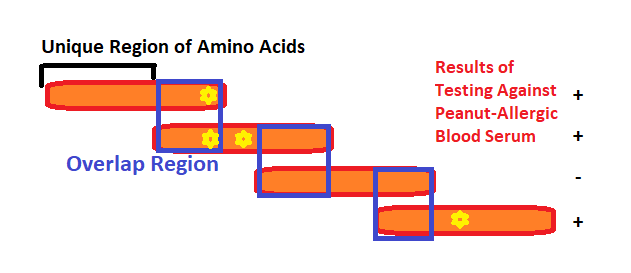
Figure 24 – a schematic of hypothetical overlapping peptide – each orange segment is a peptide, with regions that overlap in between. Regions of high binding affinity are shown with yellow stars, indicating the presence of epitopes in those regions. The response of these hypothetical peptides to a probe with peanut-sensitive IgE-containing blood is shown on the right.
From this test, three sets of amino acid residues were recognized by serum IgE as IgE-binding – amino acids 17 to 39, 41 to 80, and 114 to 157.
From these epitope-containing regions, smaller peptides were synthesized (ten amino acids long and overlapping by two) in order to more specifically identify the IgE binding regions. In Figure 25, below, the epitopes found within the peptides are identified. Epitopes in ten of the peptides were recognized by the combined pool of serum. From there, scientists tested each individual patient’s serum against those ten peptides and determined that epitopes in peptides 3, 6, and 7 were recognized by all of the patients and are thus the immunodominant epitopes for Ara h 2. Epitopes 6 and 7 both contain the amino acid sequence DPYSP, indicating that this particular sequence is salient to IgE-binding.
In the figure, each epitope region is boxed in red within its corresponding peptide. Some epitopes extend across the entire peptide, others do not. The epitopes that responded universally to peanut-allergic serum are boxed in yellow and the consensus sequence between 6 and 7 is boxed in purple. All epitopes are contextualized within the original IgE-binding regions from the first set of overlapping peptides, as they were denoted above.
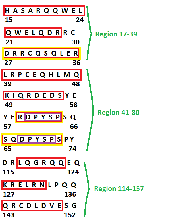
Figure 25 – Ara h 2 epitope sequences; all epitope (high IgE binding) regions in red, those epitopes with universal responses to peanut-sensitive IgE in yellow and epitope consensus sequence in purple
Recent Comments