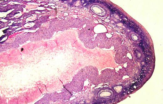
Lactating mammary gland from a cat.
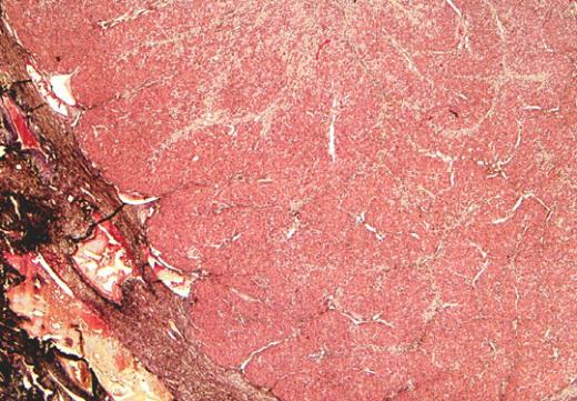
The mammary gland is a compound tubuloalveolar gland. The secretory parenchyma is divided into lobes and then lobules by connective
ViewThe mammary gland is a compound tubuloalveolar gland. The secretory parenchyma is divided into lobes and then lobules by connective
ViewThe epithelium of the oviduct mucosa contains two major cell types, ciliated and secretory. The ciliated cells are columnar in
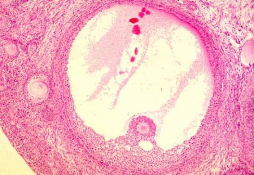
ViewThis low power micrograph shows part of a corpus luteum. The atrium and the fluid inside has disappeared as the
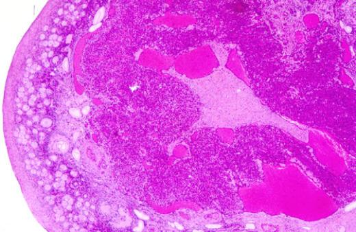
ViewThis is a low power micrograph of an ovary which contains a recently formed corpus luteum. The Graafian follicle, prior
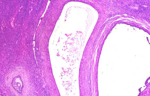
ViewIn this low power picture of the ovary (8x) you can see the relative sizes of two mature secondary follicles
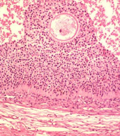
ViewIn the antrum of this secondary follicle the follicular liquor formed a precipitate and stained eosinophilic. The oocyte has reached
ViewThe antrum of this secondary follicle is quite large. Some of the proteins in the follicular liquor stained light pink.
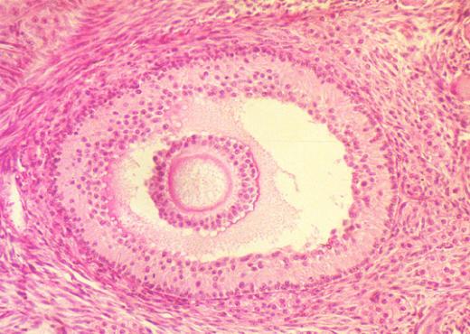
ViewAs the secondary follicle grows under the influence of FSH and increases in size, fluid (follicular liquor) accumulates between the
ViewAs the secondary follicle grows under the influence of FSH and increases in size, fluid (follicular liquor) accumulates between the
ViewAs the follicle develops, the oocyte becomes larger. The follicular cells proliferate and the oocyte is surrounded by stratified polyhedral
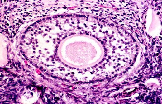
ViewThe primary follicle contains an oocyte surrounded by a single layer of cuboidal epithelial cells which sit on a basal
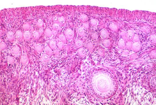
ViewBeneath the surface epithelium of the ovary is a thick layer of dense connective tissue, the tunica albuginea. The primordial
ViewThis is a low power micrograph of the surface of a feline ovary. The ovary is divided into a cortex
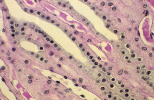
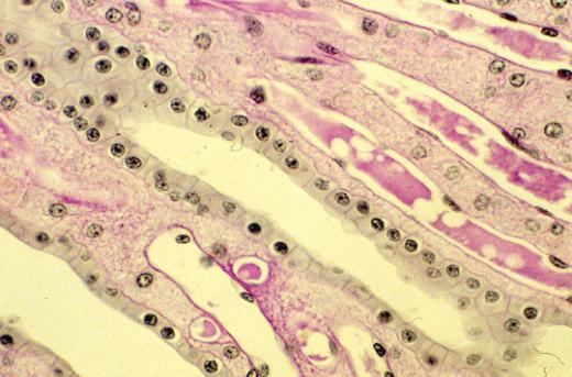
ViewIn a longitudinal section, the cuboidal nature of the cells in the collecrting duct is quite obvious. The other features
ViewIn a longitudinal section, the cuboidal nature of the cells in the collecrting duct is quite obvious. The other features
ViewThis slide was stained with PAS-hemotoxylin. Note the bright staining of the basal lamina surrounding the tubules as well as
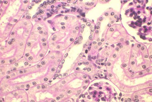
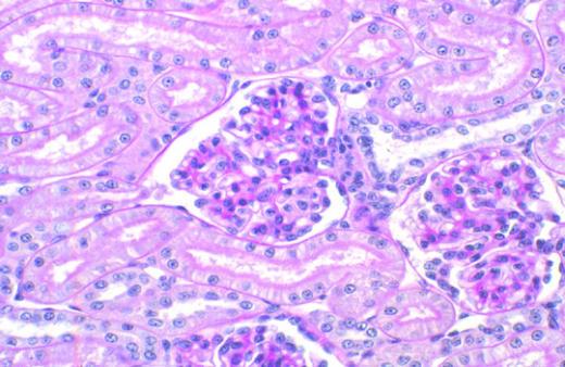
ViewIn one of the two renal corpuscles in this section you can see both the vascular pole and the urinary
ViewAnother tangential section of the respiratory epithelium gives the impression that this protion of the epithelium contains many layers of
ViewA tangential section of the respiratory epithelium gives the impression that this protion of the epithelium contains many layers of
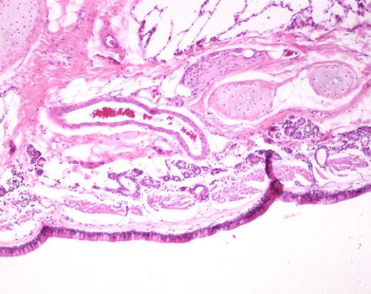
ViewAll along the respiratory tract you will find various aggregated of lymphoid tissues. These form the B. A. L. T.,
ViewThis is a micrograph of an inrapulmonary bronchus (cartilage islands, muscularis mucosae, submucosal glands). There are several bronchial blood vessels
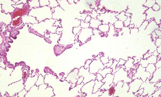
ViewThe picture can be divided into three parts. On the right is a portion of a bronchiole with its associated
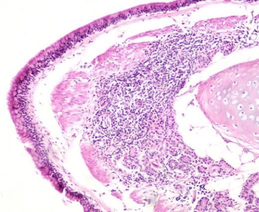
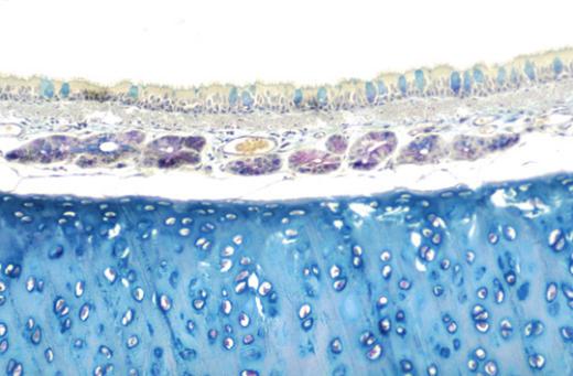
ViewThis is from the trachea of a cat at medium power. The globules of mucus in the goblet cells stain
ViewThis is from the trachea of a cat at low power. The mucus in the goblet cells stain light blue.
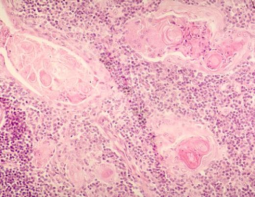
ViewThe medulla of the thymus is paler in staining when compared to that of the cortex. There are plasma cells,
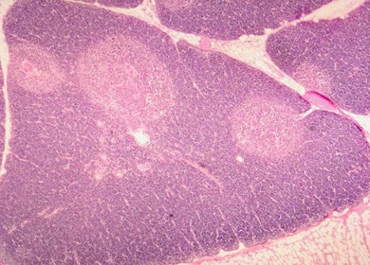
ViewThe thymus is divided into lobes and then lobules. The cortex stains darker because the lymphocytes there are closely packed
View