Urothelium 2
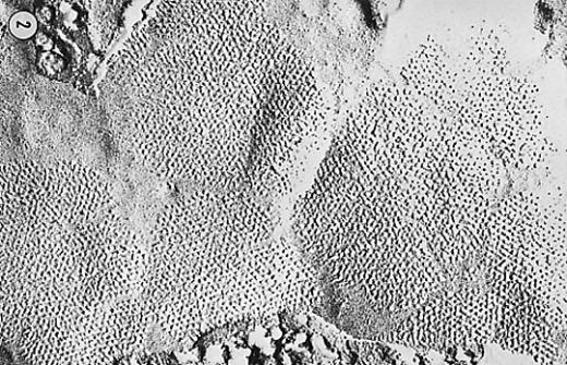
Urothelium 2 Free-fracture of luminal membrane of canine bladder urothelium showing polygonal plaques of hexagonally packed particles. The protein that
ViewUrothelium 2 Free-fracture of luminal membrane of canine bladder urothelium showing polygonal plaques of hexagonally packed particles. The protein that
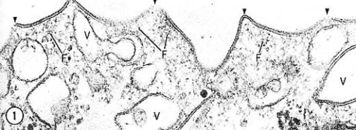
ViewUrothelium 1 Thin section electron micrograph of luminal plasma membrane of superficial cell of dog urinary bladder. The outer leaflet
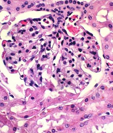
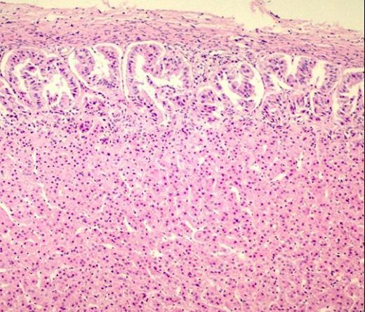
ViewIn this renal corpuscle you can see both the vascular and the urinary poles. Some of the capillaries in the
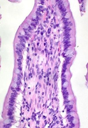
ViewTip of a villus from the jejunum of a dog. There is some separation artifact at the junction between the
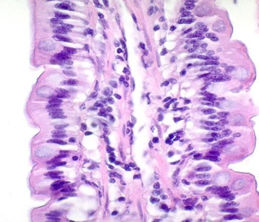
ViewCental lacteal in the core of a villus. The lacteal is part a blind-ended lymphatic vessel and is involved in
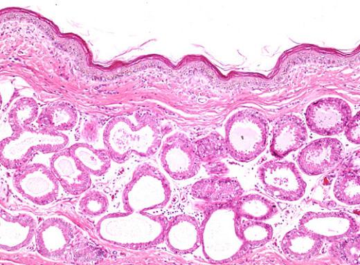
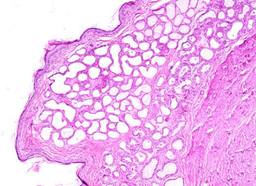
ViewIn the dog, the glands of the anal sac (sinus paranales) are large aprocrine sweat glands. They are prone to
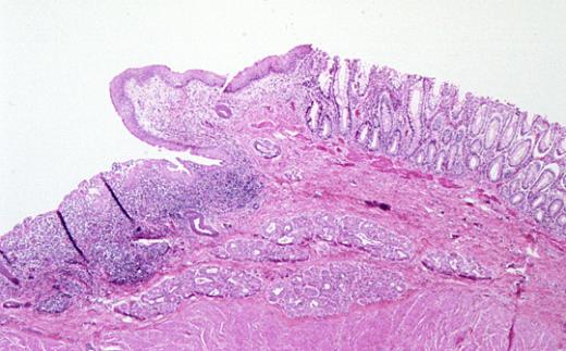
ViewThis low magnification micrograph shows the anal canal area of a dog. A variety of glands are included here but
ViewGlands of the anal sac from a dog. PK1636. J11a. H&E. Original mag. 10x. Anal sac. Glands of anal sac.
ViewRecto-anal junction in the dog. The rectal area is covered by colonic mucosa. The anal canal is covered by stratified
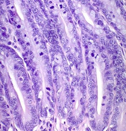
ViewThe epithelium of the GI tract is replaced on a regular basis. In the intestine mitosis takes place in the
ViewVilli from the jejunum of a retriever. PK0576. H&E. Original mag. 33x. Jejunum. Samll intestine. Digestive System. Dog.
ViewJejunum of a retriever at low magnification. Note the villi and the crypts. PK0574. H&E. Original mag. 13x. Jejunum. Small
ViewTip of villi from the duodenum of a dog. The mucus in the goblet cells stained brightly. The lamina proria
ViewThis section was stained with PAS. Notice the mucus in the goblet cells stained differently from that of the Brunner’s
ViewAt this magnification you can clearly see the cells that make up the Brunner’s glands. Notice the size, shape and
ViewA medium power micrograph of the duodenum from a dog. J8. PK1624 PTAH. Original mag. 25x. Brunner’s glands. Duodenum. Small
ViewDuodenum at low power showing the villi, the crypts and the Brunner’s glands. Note the location of the muscularis mucosae.
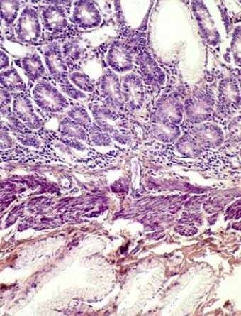
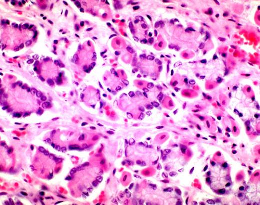
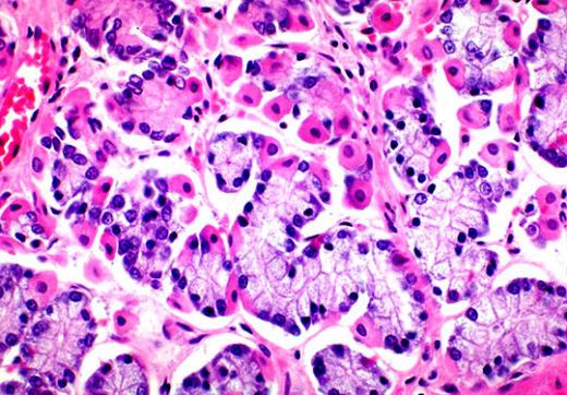
ViewThe parietal cells of the fundic stomach stain eosinophilic due to the large number of mitochondria in the cytoplasm. These
ViewMucous glands in the submucosa of the esophagus in a dog. In the dog these glands are found throughout the
ViewSinus or tactile hair follicle is specialized for tactile sense. Each is a single follicle which is much larger than
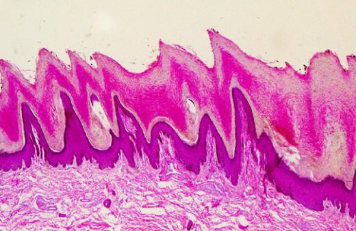
ViewThe digital pad of the dog contains thick stratified squamous keratinized epithelium. The cellular layers of the epidermis and the
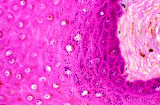
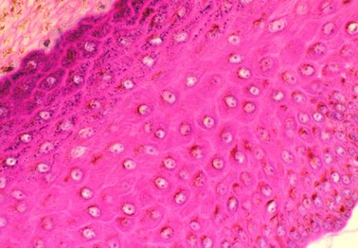
ViewThis section shows the stratum spinosum, the stratum granulosum and the stratum lucidum of the thick skin in the foot
ViewThis micrograph spans most of the layers of the thick skin. In one corner is the stratum basalis while in
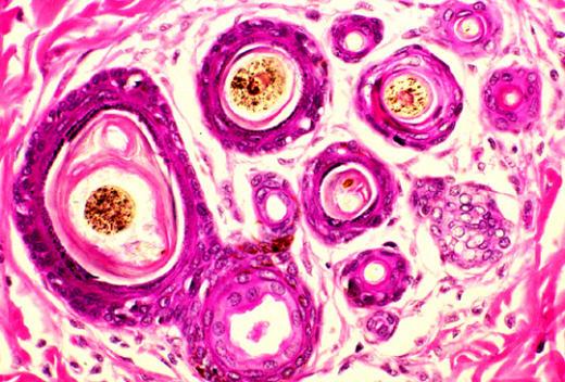
ViewSeveral hair follicles cluster together in the dermis to form compound hair follicle. Usually there is a primary hair follicle
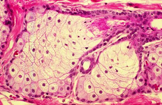
ViewThe aprocrine sweat glands have a secretory portion and a ductal portion. The epithelium of the secretory portion contains cuboidal
ViewThe sebacous glands secrete an oily substance, sebum, which acts as an antibacterial agent and, in hairly mammals, as a
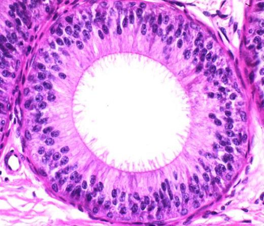
ViewHere you can make out the seterocilia. However, it may be difficult to tell exactly what these structures are made
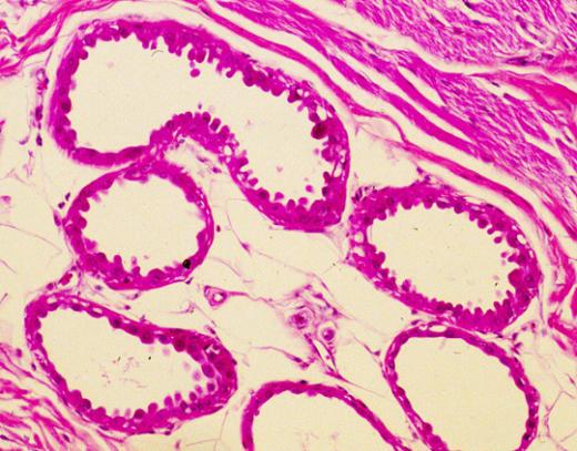
ViewLow power micrograph of epididymis from a dog. This is a long tubular structure that is highly coiled and compact.
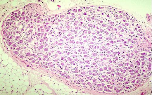
ViewOrgan: Testis, Tissue: Leydig cells, Species: Dog. Comments: A cluster of interstitial cells of Leydig is surrounded by seminiferous tubules
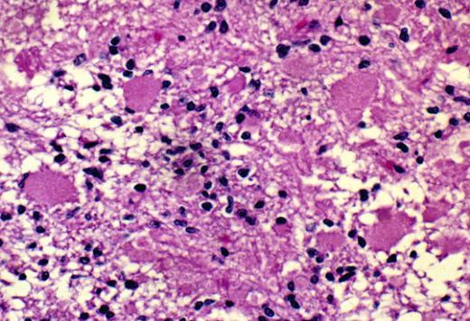
ViewOrgan: Adrenal, Tissue: Autonomic ganglion, Species: Dog. Comments: Parasympathetic ganglion with numerous basophilic neuronal cells in the adrenal of a
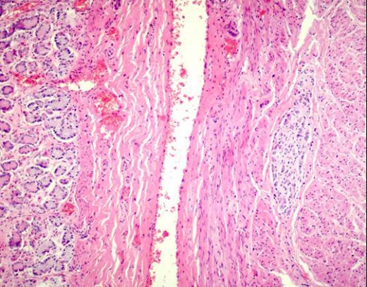
ViewOrgan: Stomach, Tissue: Myenteric plexus of Auerbach, Species: Dog. Comments: The myenteric plexus of Auerbach is found between the two
ViewOrgan: Stomach, Tissue: Myenteric plexus of Auerbach, Species: Dog. Comments: The myenteric plexus of Auerbach is found between the two
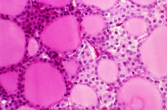
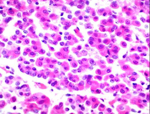
ViewThe calcitonin secreting C-cells (parafollicular) cells have pale cytoplasm and are found between the follicules. Note the amount of cytoplasm
ViewThis section contains the pars distalis, the pars intermedia, reamins of Rathke’s pouch, and the pars nervosa. The pars distalis
ViewThe nuclei seen in this section of the neurohypophysis belong to the supportive cells. The cell bodies of the unmyelineated
ViewThis section contains the pars distalis, the pars intermedia, reamins of Rathke’s pouch, and the pars nervosa. The pars distalis
ViewThe cells of the zona reticularis produce small amounts of sex hormones. Being steroid synthesising and metabolizing cells, they have
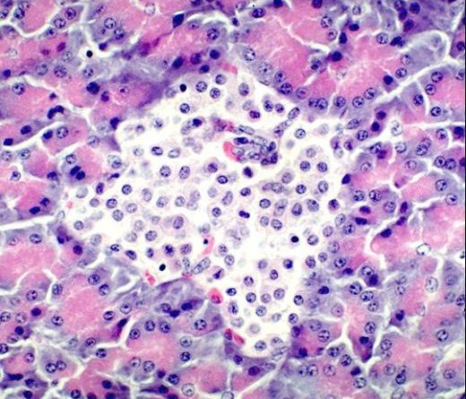
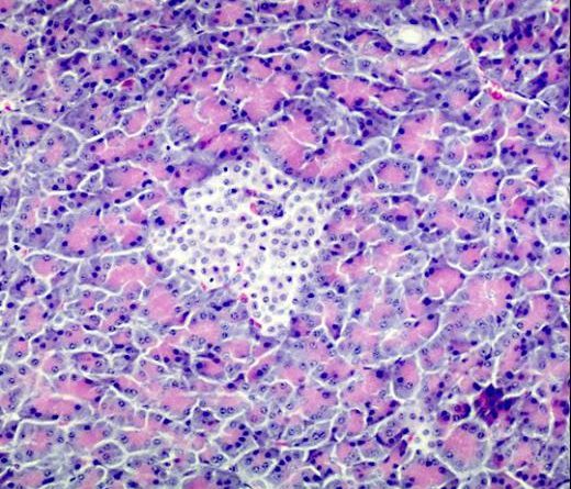
ViewOrgan: Pancreas, Tissue: Islet of Langerhans, Species: Dog. Comments: Several secretory cell types, plus a network of capillaries makes up
ViewOrgan: Pancreas, Tissue: Islet of Langerhans, Species: Dog. Comments: Islets of Langerhans are endocrine structures that occur as clusters of
ViewOrgan: Pituitary, Tissue: Pars distalis, Species: Dog. Comments: A few chromophobes are found among the numerous acidophils.
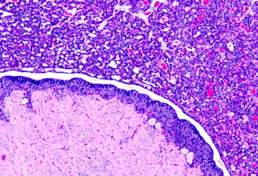
ViewSystem: Endocrine, Organ: Adrenal, Tissue: Cortex. Species: Dog. Comments: Low power micrograph of dog adrenal cortex including the zona arcuata
ViewOrgan: Pituitary, Tissue: Pars distalis, Species: Dog. Comments: The majority of the cells in this micrograph are acidophils. The rest
ViewOrgan: Adrenal, Tissue: Cortex, Species: Dog. Comments: Adrenal cortex with capsule. The cells in the outermost zone of the cortex
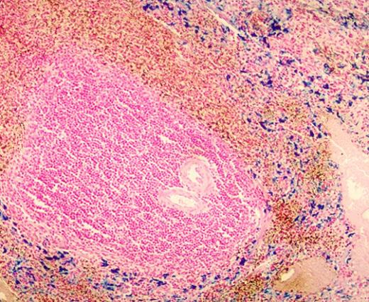
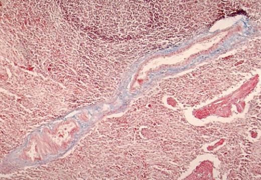
ViewThis section was stained by the Gomori iron method in which the metal stained blue. The nuclear counter stain used
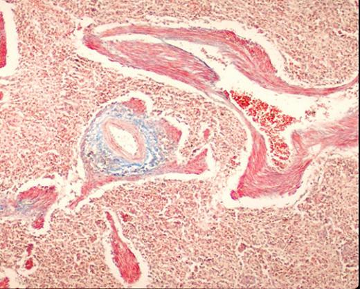
ViewThe trabecular artery has a distinctive tunica media made up of smooth muscle cells. It also has a tunica adventitia
ViewThe trabecular vein in the spleen has an tunica intima made up of a layer of endothelial cells. The rest
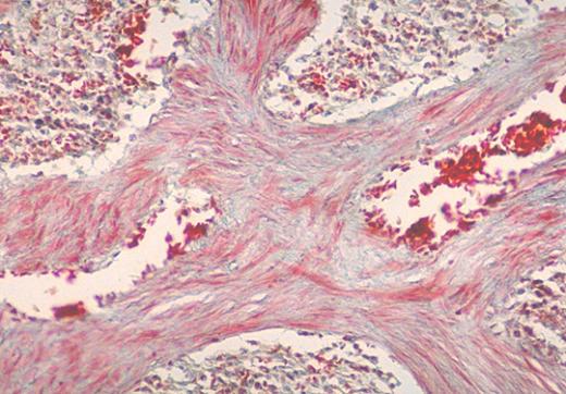
ViewThe trabeculae in the spleen contain smooth muscle cells and connective tissue. The trabecular artery has its own muscular wall
ViewThis slide shows the longitudinal section of a trabecular artery in the spllen. Notice the smooth muscle cells that make
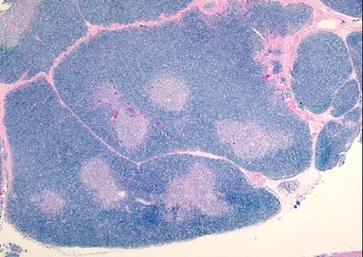
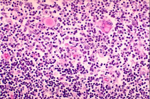
ViewSystem: Lymphoid, Organ: Thymus, Tissue: Cortex, Species: Dog. Comments: The cortex contains tightly packed small lymphocytes that make this area
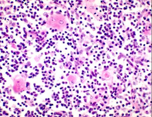
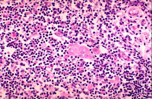
ViewSystem: Lymphoid, Organ: Thymus, Tissue: Medulla, Species: Dog. Comments: The eosinophilic Hassall’s corpuscles are characteristic features of the thymus. The
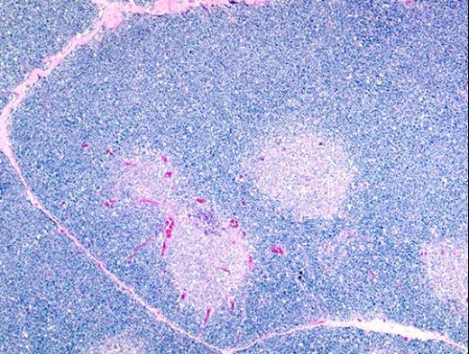
ViewSystem: Lymphoid, Organ: Thymus, Species: Dog. Comments: One half of the micrograph shows the cortex with tightly packed small lymphocytes
ViewThe cortex stains darker than the medulla because the former is mostly made up of densely packed lymphocytes. The cortex
ViewA thin connective tissue forms the capsule of this organ. Connective tissue septae extending inward from the capsule divide the
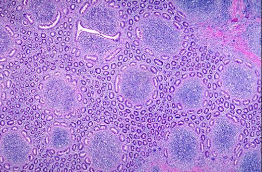
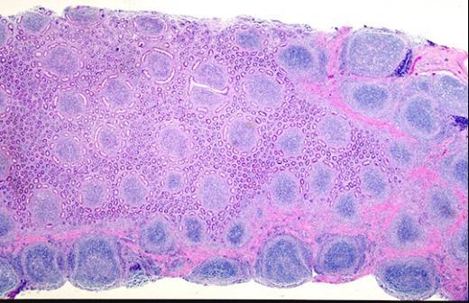
ViewSystem: Lymphoid, Organ: Ileum, Tissue: Peyer’s patch, Species: Dog. Comments: Peyer’s patch cut “en face”. As the cut slants deeper
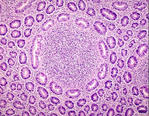
ViewSystem: Lymphoid, Organ: Ileum, Tissue: Peyer’s patch, Species: Dog. Comments: Peyer’s patch cut “en face”. Crypts of Lieberkuhn surround a
ViewSystem: Lymphoid, Organ: Ileum, Tissue: Peyer’s patch, Species: Dog. Comments: Peyer’s patch cut “en face”. Crypts of Lieberkuhn surround the
ViewSystem: Lymphoid, Organ: Ileum, Tissue: Peyer’s patch, Species: Dog. Comments: Low magnification of the Peyer’s patch cut “en face”. The
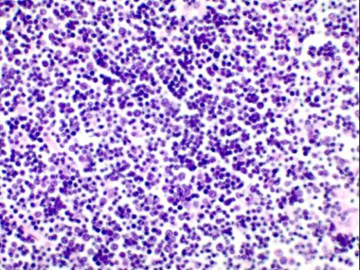
ViewOrgan: Thymus, Tissue: Medulla, Species: Dog. Comments: In the medulla of the thymus the cells are further apart. Thus the
ViewOrgan: Thymus, Tissue: Medulla, Species: Dog. Comments: In the medulla of the thymus the cells are further apart and the
ViewIn the cortex of the thymus illions of lymphocytes are produced here each day and these are, by definition, T
View