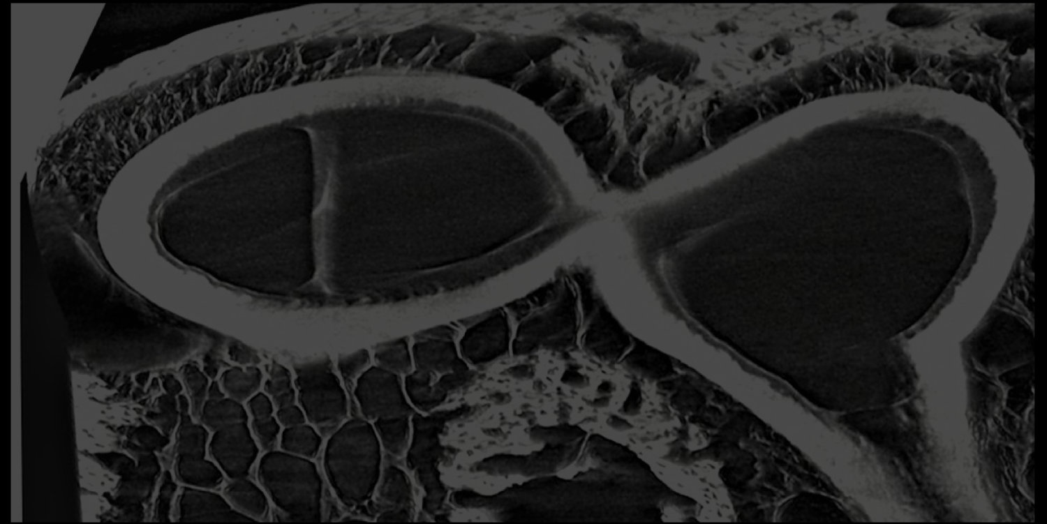
Non-Uniformity In The Periodontal Ligament

The periodontal ligament (PDL) is the only soft tissue in the entire complex of the teeth and jaw bones, and therefore has a major role in controlling tooth movement and bone remodeling. Our research show that the 3D structure of the collagen network within the PDL is not uniform. We identified a unique region that we named the “Dense Collar” due to its high collagenous density and stiffness. This project is aimed at studying the role of the “Dense Collar” in tooth function and how it can be structurally manipulated to treat periodontal disease and facilitate orthodontic tooth movement. To do that we use mechanical testing, Mass spectrometry, Raman, MicroCT, AFM, molecular and cell biology, multiphoton microscopy and different image analysis tools and algorithms.
The Function of Blood Vessels in the PDL and Bone Remodeling

Differently from most ligaments, the periodontal ligament has a rich network of blood vessels. The remodeling process of the alveolar bone is closely related to the blood supply to the PDL. The principal notion in orthodontics is that compression in the periodontal ligament creates ischemic events which trigger recruitment of osteoclasts and bone resorption. However the effect of the force levels and duration on the resorption rate and extent are not well understood. In order to elucidate the function of the blood vessels in the PDL we are using transgenic mice and a new optical imaging method that enables us to unravel the intriguing role of blood vessels in the alveolar bone remodeling process. For this purpose we use tissue clearing, multiphoton microscopy, machine learning and image analysis.