Rapid contracting and low, flexible pricing
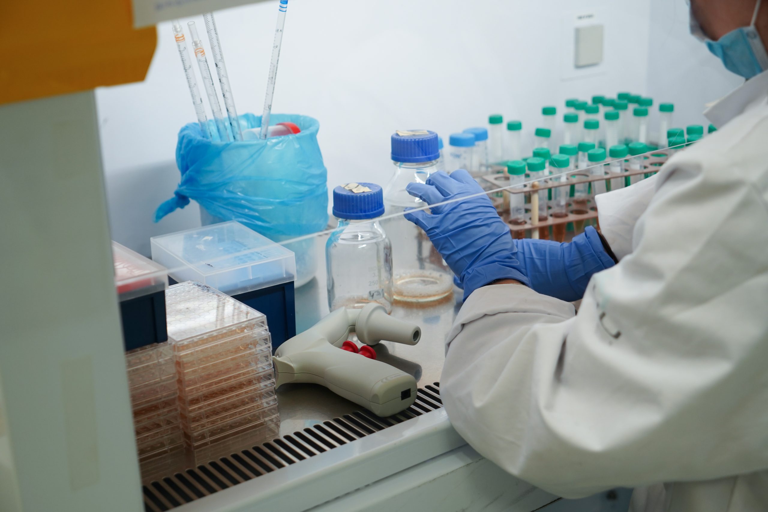
Tufts Comparative Medicine Services (CMS) provides an array of in vivo services to support preclinical and biomedical research to industry and to academic research laboratories in downtown Boston, the New England area and beyond. Being one of the first academic Contract Research Organizations (CROs) Tufts CMS has oversight and management of the AAALAC accredited animal care and use program on the Boston campus. Leading the way in contract preclinical and biomedical research, Tufts CMS vivariums and teams provide resources for academics, bio tech from large pharma to startups and more with rapid contracting and low, flexible pricing.
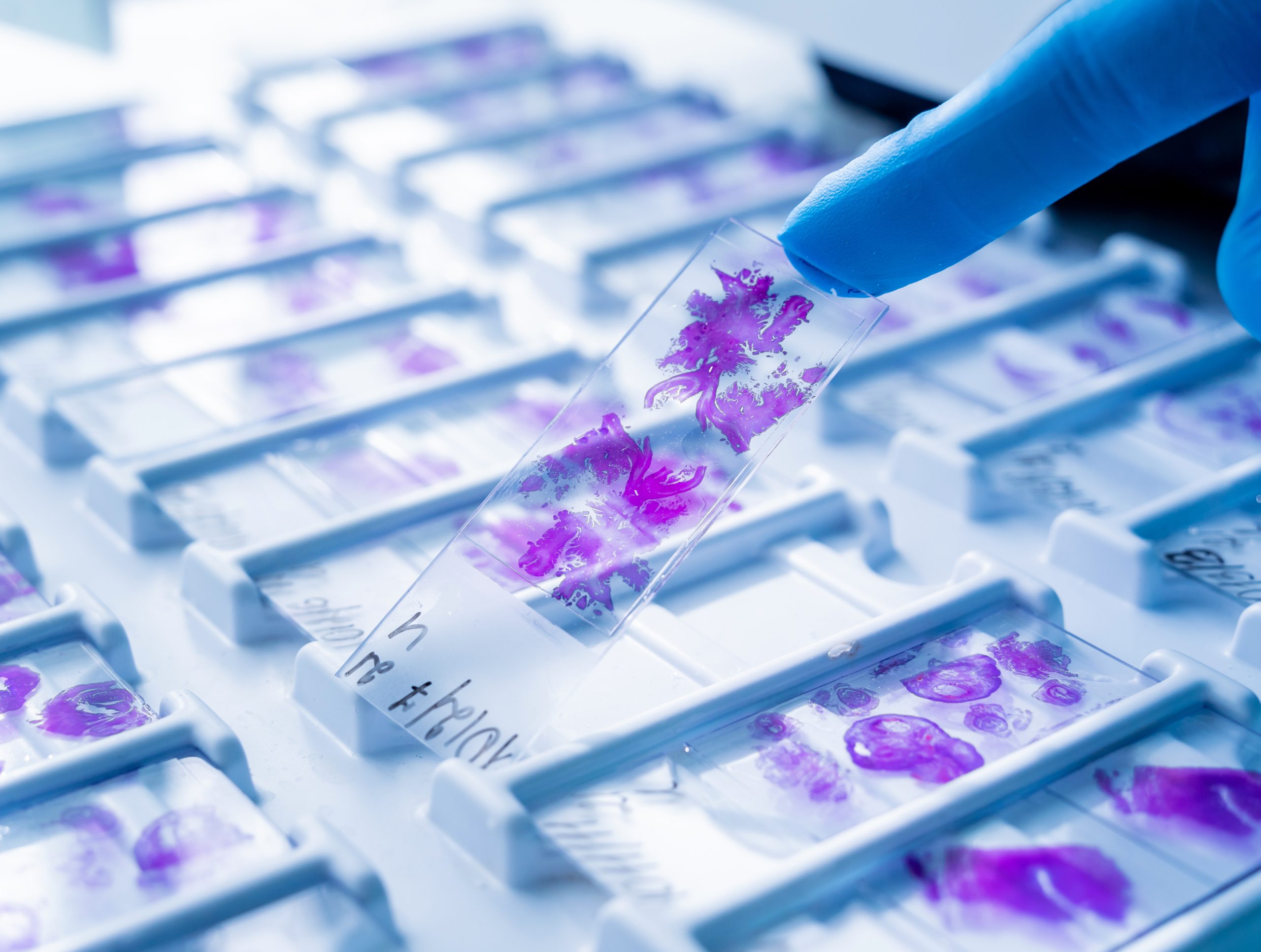
laboratory offering clinical and anatomic pathology services. CPS services offered include: hematology (complete blood counts with white blood cell differentials, serum biochemical panels); parasitology; microbiology (bacterial and fungal identification, antimicrobial sensitivity); analysis of urine and other body fluids; serology for numerous bacterial, viral and parasitic infectious agents; and environmental monitoring. All tests are performed by qualified laboratory professionals trained in veterinary laboratory techniques. The CPS also has access to a network of reference laboratories to provide tests that are not performed in-house. CLICK HERE FOR MORE INFO
Preclinical imaging involves the use of advanced imaging modalities such as MRI (Magnetic Resonance Imaging), PET (Positron Emission Tomography), CT (Computed Tomography), ultrasound, and optical imaging to study biological processes in animal models before clinical trials in humans. This non-invasive approach provides detailed visualization of the anatomy, function, and molecular characteristics of tissues and organs in vivo. Preclinical imaging aims to understand disease mechanisms, evaluate the efficacy and safety of new therapeutic interventions, and facilitate the translation of scientific discoveries from the laboratory to clinical applications. It is essential for drug development, biomarker discovery, and the advancement of personalized medicine.
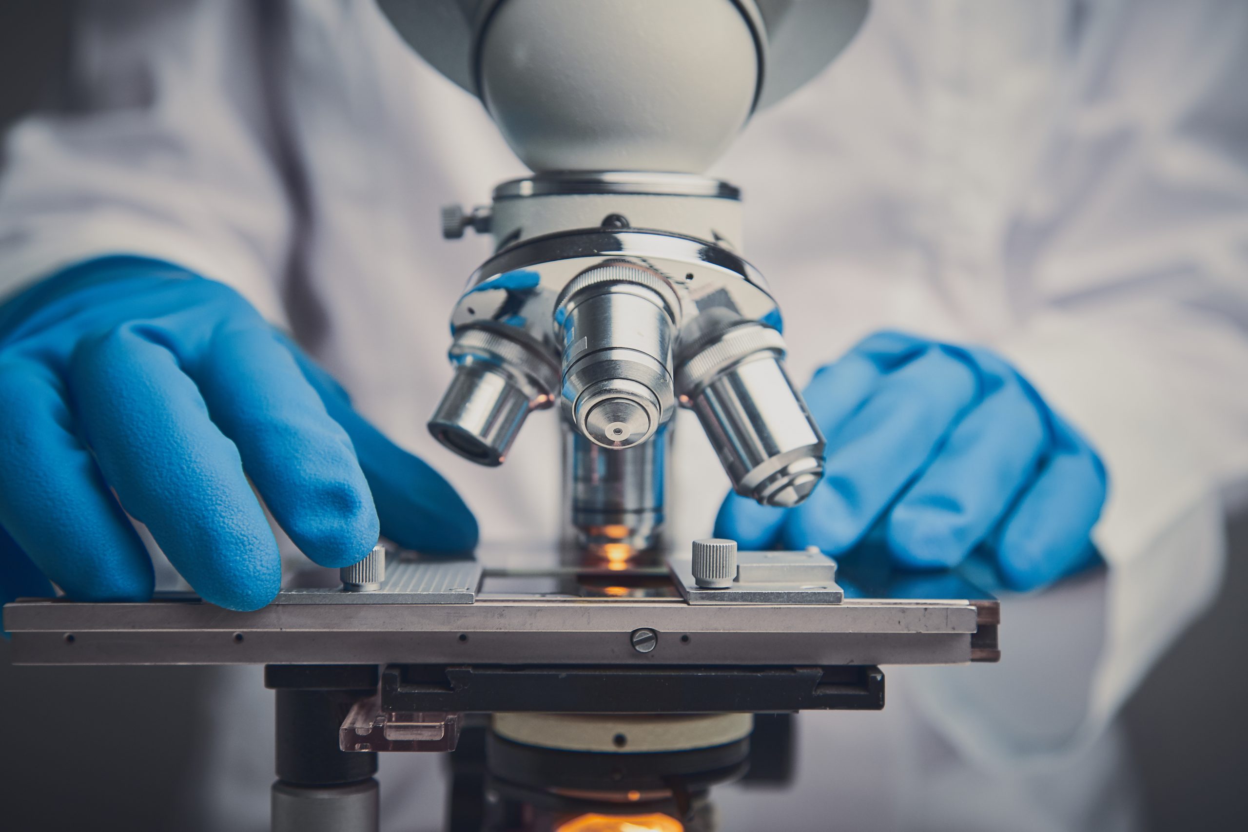
In vitro imaging, on the other hand, focuses on the study of biological samples outside their natural biological context, in a controlled laboratory setting. Techniques such as fluorescence microscopy, confocal microscopy, electron microscopy, and various optical imaging methods are used to examine cells, tissues, or biochemical substances in isolation. This allows researchers to visualize cellular structures, track molecular interactions, study cell behavior, and understand disease mechanisms with high resolution. In vitro imaging is crucial for basic research, drug discovery, and the development of diagnostic methods, providing detailed insights that inform in vivo studies and clinical applications.
Together, preclinical and in vitro imaging offer a comprehensive approach to studying biological processes, bridging the gap between laboratory research and clinical practice.
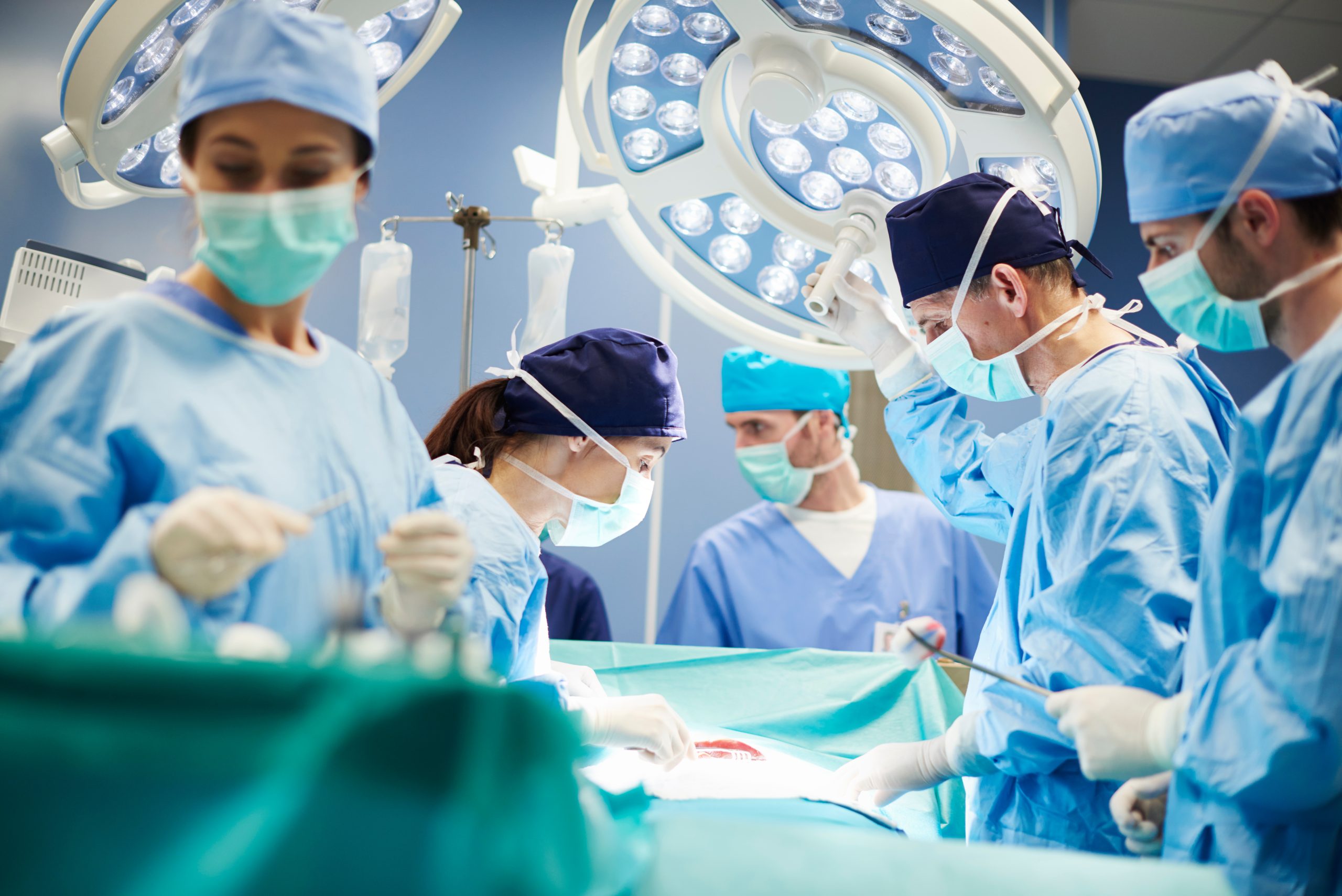
Preclinical Services provides the facilities and technical assistance needed to support research procedures. PS can be hired in a number of capacities depending on the desired scope of work. PS can perform a single task or conduct an entire study including collection and processing of data and results. The PS team provides services for a variety of routine in vivo testing, including minor and major surgeries, anesthetic support, drug administration, collection of tissues, and euthanasia of animals. PS assistance includes, but is not limited to: pharmacokinetics, tumor studies, efficacy studies, immunogenicity studies, toxicity studies, and a variety of other special models as requested by clients. CLICK HERE FOR MORE INFO
Preclinical Services provides the facilities and technical assistance needed to support research procedures. PS can be hired in a number of capacities depending on the desired scope of work. PS can perform a single task or conduct an entire study including collection and processing of data and results. The PS team provides services for a variety of routine in vivo testing, including minor and major surgeries, anesthetic support, drug administration, collection of tissues, and euthanasia of animals. PS assistance includes, but is not limited to: pharmacokinetics, tumor studies, efficacy studies, immunogenicity studies, toxicity studies, and a variety of other special models as requested by clients. CLICK HERE FOR MORE INFO