Guest Post by Ila Anand, Micro
A startling number of viral epidemics have made major media headlines in recent years. In 2014, the Middle Eastern Respiratory syndrome coronavirus (MERS-CoV) was quickly brought to America’s attention after two reported cases in Indiana and Florida. 2015 was the year the world went into Ebola frenzy and U.S. hospitals took extreme precautions to manage suspected infected patients. This year, the Zika virus has caught the CDC’s eye and for good reason. As Zika continues to spread from Brazil through the Americas, its arrival in the U.S. this summer is inevitable. Although no vector-borne cases have been reported inside the U.S. yet, over 150 travel-associated cases have been reported. Public health departments across the U.S. should brace for the next likely step: the moment when Zika passes from traveler-infected blood to a local mosquito and then to another person.
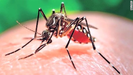
The Zika virus was first isolated in Uganda in 1947 from the Zika Forest, where researchers from the Rockefeller Foundation were studying yellow fever. These researchers experimentally used rhesus monkeys that were set out in cages in treetops as bait for mosquitos carrying yellow fever virus. Ironically, instead of yielding yellow fever virus from the blood of these monkeys, the researchers discovered Zika and speculated the virus had been lurking chronically in African monkeys for millennia. The virus was later isolated from mosquitoes of the Aedes genus in the same Zika forest and Aedes has since been identified as the vector of Zika. Eventually, Zika virus was discovered to infect humans across the African continent as well as in South Asia and Southeast Asia. More recently, circa April 2015, the virus has spread to the South America.
In humans, the virus manifests infection known as Zika fever, which often produces no symptoms to mild symptoms, such as headache, fever, rash, bloodshot eyes, and joint pain. Recently, the spread of Zika in South America has been linked to the growing number of infants born with microcephaly in Brazil. Microcephaly is a neurological condition in which the brain and skull fail to grow at a normal pace, resulting in a significantly smaller head size. At first the link to Zika was purely correlation, however, a recent report published in Cell Stem Cell directly demonstrated that Zika is able to infect and kill lab-cultured human neural progenitor cells. These neural progenitor cells were derived from induced pluripotent stem cells (iPSCs) and scientists tested Zika’s “tropism” by comparing percent infection across four cell types: neural progenitor cells, immature neurons, embryonic stem cells, and human iPSCs. While less than 20% of iPSCs, embryonic stem cells and neurons became infected, up to 90% of neural progenitor cells contained the virus and Zika either killed these cells or slowed their proliferation significantly. These findings may begin to unearth some possible mechanisms to how Zika infects and damages fetal brain tissue. Since neural progenitor cells give rise to a larger population of neurons and glial cells in the brain, infection of these cells could impact the neurons they produce and possibly affect brain development. In addition to microcephaly, Zika has also been linked Guillain-Barré syndrome, a sickness in which the person’s own immune system damages nerve cells, causing muscle weakness and sometimes paralysis. However, clinical findings from Brazil are still preliminary and there’s a need for more compelling evidence.
Sources –
- History of Zika, National Geographic
- H. Tang et al., “Zika virus infects human cortical neural precursors and attenuates their growth,” Cell Stem Cell, doi:10.1016/j.stem.2016.02.016, 2016
- Center for Disease Control (CDC) Zika information
- Zika and Guillain-Barre Syndrome, Time Magazine

