Category Archives: Techniques

Top Techniques: Confocal Microscopy

by Alenka Lovy

What is confocal microscopy?

Reading and thinking about cell biology is very interesting no doubt, but I find that to be able to see biological processes by live microscopy just amplifies the questions at hand so much! Have you ever seen movies of cells dividing? I remember when I first did. It was hard to go from the picture perfect diagrams of the textbook to the real thing, but after a few times of watching the movies I saw the perfect (or not so perfect) progression through all the steps. Maybe it was the timing of it, or just being able to see the tangle of chromosomes trying to line up, and then the sudden division, I found it so breathtaking! Live cell microscopy has been my tool of preference to answer many biological research questions ever since.

Confocal microscopes in particular are powerful because they optically slice through a specimen (even live cells) and allow 3D image reconstruction in up to four different fluorescent channels. Confocals are built to scan point by point through your sample using laser light, and image just one particular plane of focus. This is very different to the standard fluorescent microscope which illuminates and images the entire sample at once, including out-of-focus light. The confocal is used to obtain clearer images of subcellular details that cannot be imaged with the fluorescent microscope and is especially useful for co-localization studies. There are many exciting techniques you can use with the confocal including fluorescence recovery after photobleaching (FRAP) with which you can observe protein mobility and recovery, fluorescence resonance energy transfer (FRET) which can show protein interactions, as well as photoactivation/uncaging studies.


BOX 1: What confocal can do for you (and your mitochondria)

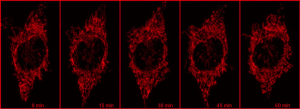
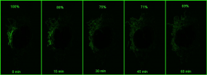
Using a photoactivatable GFP targeted to the mitochondria to measure mitochondrial fusion is a nice demonstration of the precision and quantitation that can be achieved using a confocal. Mitochondria are amazingly motile and networked and look like spaghetti . They also undergo constant fission and fusion, which can be difficult to capture. The top panel in the figure below shows the mitochondrial network (z stack) labeled with TMRE in Hela cells imaged every 15 min for 1 hr. It is impossible to capture which mitochondria are fusing.

red

green

However, if a small portion of the network is photoactivatedand then imaged in z stacks over time, the signal can be monitored over time (bottom panel in the above figure). As the mitochondria fuse, the GFP protein becomes diluted in the larger volume of the network that has not been photoactivated, and the extent of dilution can be quantified and used as a measure of mitochondrial fusion.


What facilities does Tufts have for confocal microscopy and other imaging techniques?

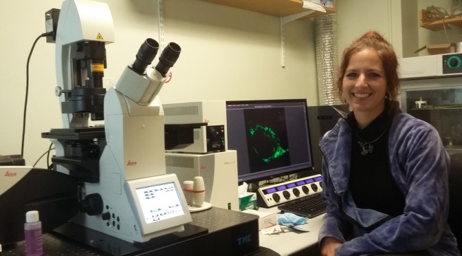
The Tufts Imaging Facility has four confocal microscopes and most are equipped with the standard 405nm, 488nm, 561nm and 633nm laser lines, which is important to know when choosing fluorophores. Using the Fluorescence SpectraViewer online will help you determine if the emission spectra of your fluorophores overlap such that crosstalk between them can be minimized. We have two inverted microscopes equipped for live cell imaging, and two upright microscopes that are usually used for fixed samples and 3D reconstructions. While imaging living cells, you can use an automated focusing mechanism which employs an infrared laser that keeps track of the coverslip, and therefore your sample. If you’ve ever had to adjust the focus yourself over several hours, you know just how powerful this feature is!

The Nikon A1R inverted confocal has a resonant scanner capable of high speed imaging (500 frames/sec at 512×512 pixel resolution) suitable for ion imaging and is being used for calcium imaging in cardiomyocytes. It also comes in handy during very long tiled scans with z-stacks, and although the image quality is slightly sacrificed, depending on the resolution needed, the gain in speed may well be worth it.

The Leica SP8 inverted confocal has a HyD sensitive detector and can be used with very low laser powers allowing longer imaging of easily bleached samples. For example, measuring how quickly a photoactivated GFP spreads within the mitochondrial network every minute over an hour would bleach the signal before all the information was collected on a regular detector compared to a HyD detector.

Another good technique to avoid bleaching in live cell imaging is to use the total internal reflection microscope (TIRFM). On this microscope, you can adjust the angle of laser light with which you illuminate your sample. There is a particular angle at which all the laser light is internally reflected, except for a 100nm evanescent wave. With this you can then image processes close to the membrane, such as receptor insertion/cycling. Very often you can image a little bit deeper than the 100nm, and because the laser is at an angle, you will not bleach your specimen as fast. As opposed to confocal, the TIRF system has a sensitive EMCCD camera, enabling faster imaging (I have been looking at calcium sparks at 50 ms/frame).

The Leica upright microscope has water immersion objectives that have a large working distance and work well for thick cleared samples such as mouse brains or zebrafish embryos.

Finally, in addition to standard fluorescent microscopes, we also have the automated Keyence fluorescence microscope which can scan up to 3 slides and stitch large images together in four channels as well as in brightfield. If large tiled scans are needed, this may be the instrument of choice due to the speed and ease of use.

For more information about the instruments in the Tufts Imaging Facility, please visit our website. If you would like to use an instrument or need help planning an experiment please email me at:

Alenka.Lovy@tufts.edu.

If you’d like to learn more about microscopy in general, the Molecular Expressions Microscopy Primer is a great resource.

Techniques – Go with the Flow

What is Flow Cytometry and what can it do for you?

by Stephen Kwok & Allen Parmelee

Flow Cytometry is something I never heard about in school, but once I learned about it, the possibilities seemed endless as to how I could use it as a tool to make work and research better. FACS (Fluorescence Activated Cell Sorting) Sounds like an office tool, not a state of the art piece of scientific equipment. In reality, it is like a multitude of fluorescent microscopes all working together to gather data at the same time. Wait, it gets better…you can actually physically separate your cells from one single cell per well on a 96 well plate, to millions of cells in a 15ml tube! The human eye has a habit to have bias; these machines convert the analog data into a digital plot or histogram that can’t be argued with! Is it 30% positive or 35% positive? Yes, we can actually tell the difference!

Let’s back up a step here. The technology is best used if you have markers for your cells. You can take fluorescently labeled antiboties to identify cells. Let’s say you are looking for stem cells. Cd34, SCA-1, and c-Kit are common for hematopoietic stem cells. Label these three, throw in a viability marker, and you have successfully identified these cells. You can move forward with your experiment and simply ANALYZE the cells. Or, you can try to isolate these cells by SORTING them. Fluorescent protein transfections with a GFP or RFP marker are common. Why grow cells in harsh selection media when you can simply pluck them out and put them into a plate? I need to do some PCR, but I have to figure out how to get 1 cell, 5 cells, 25 cells, 50 cells. Limited dilution is going to take me forever! In as fast as 30 seconds you can have those exact numbers of cells lined up into your pcr tubes or a 96 well plate.

At our facility we have cell analyzers available for use 24/7. We train people in basic theory, and then help them get started on how to run the instruments. Sorting, however, is a little more complicated and is done by the two intimidating guys running the facility: Allen and Steve. 

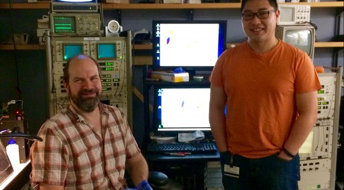
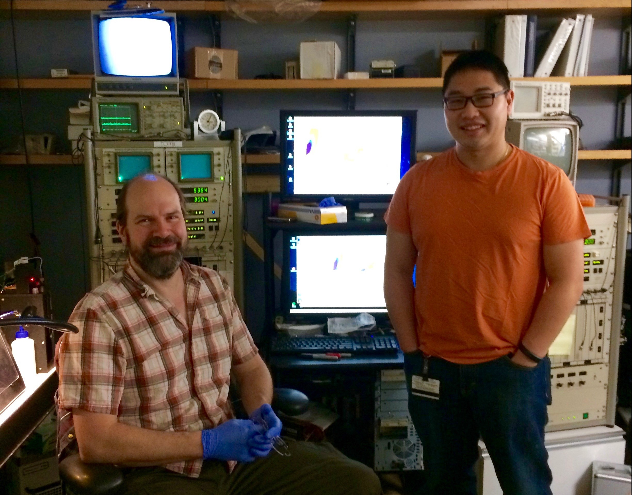
Steve & Allen, the Flow Bros (right to left)
Allen Parmelee & Stephen Kwok, the Flow bros (left to right)


There are always plenty of questions to answer about FLOW. How fast is fast? Well the Analyzers can run approximately 3,000 cells per second. The high speed cell sorters. 30,000 cells per second! This can translate to over 100e6 per hour. How sensitive are the machines? We can detect one cell in 10e6 cells! How many markers can I use? The most common is 4 different colors at a time, but we could do up to 17. Be wary, however, just because we said you can. Doesn’t mean you should. Work smarter, not harder! I have 4 different populations: can I sort them all at once? Yes! In fact, we can do up to 6 simultaneous separate populations at once.

How can I do good flow cytometry? The key is sample prep! Yes, they seem like magical boxes, but the experiment is only as good as the components. Titer your antibodies. TEST them with a positive control. Bring a negative or untreated control as a baseline. Would you run a gel without the markers? Find the correct markers, and look for the greatest separation. Cells need to be in Single Cell format. It is highly recommended to filter/strain your samples because the pathway for the cells are 70-150um in size, a clump of cells can clog the machines and render them inoperable.

Come by, check out the machines, ask us questions…we hope you’ll be pleasantly surprised at the possibilities.

Tufts Laser Cytometry, Jaharis 5th floor. 

Special points of interest:

  • No Color Cloning
  • Bulk Sorting
  • Fluorescent work
  • Cell Cycle
  • Apoptosis
  • Ca+ Flux
  • Single Cell PCR
  • Rare Event
  • Bacteria
  • CRISPR
  • Stem Cells
  • Neurons
  • Transfections
  • Infections
  •  Libraries


Notes from the North – MMCRI Transgenic Core

MMCRI transgenic core

The Maine Medical Center Research Institute (MMCRI) has partnered with Tufts to provide a professional core facility that has over 15 years of experience providing high quality services for the generation of mouse transgenic strains including the use of CRISPR/Cas, cryopreservation of mouse germ cells, and imaging, including MRI and microCT. Mice are generated in a full barrier, AAALAC-accredited animal facility in a transgenic production room that facilitates direct importation of mice into the Tufts barrier facility. Contact us to discuss your mouse and imaging projects.

Mouse Transgenesis

Source - MMCRI transgenic core
Source – MMCRI transgenic core

We provide microinjection to generate your mouse models. Services include microinjection of fertilized oocytes with traditional DNA transgenes, or microinjection of CRISPR/Cas. ES cell injection is also performed. Contact us to design your CRISPR mouse project – cost depends on type of modification, strain of mouse, and days of injection. We have a high surveillance production room that will allow importation of mice direct into some barrier facilities.

Contact – Lucy Liaw, Ph.D., liawl@mmc.org

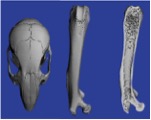
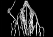
microCT

Source - MMCRI transgenic core
Source – MMCRI transgenic core

We house a Scanco high speed in vivo microCT scanner X-ray system. Our microCT facility has extensive experience in bone imaging and quantification, and can work on other projects where tissues are provided, i.e., vascular imaging of samples perfused with microfil. We provide quantification and any 3D images of the samples as required. Contact us to get a project quote. Pricing is based on hours of scanning and analysis time.

Contact – Lucy Liaw, Ph.D. liawl@mmc.org

Small Animal MRI

Source - MMCRI transgenic core
Source – MMCRI transgenic core

Our MRI facility houses a Bruker Pharmascan 7T, 300 MHz imager with 100 μm resolution. Services include anatomical imaging of most organs, angiography, proton spectroscopy and localized spectroscopy, and cardiac imaging, including diastolic and systolic dimensions of the ventricle. We can house “clean” animals at our facility for studies requiring longitudinal imaging. Contact us for mor information.

Contact – Ilka Pinz, Ph.D. pinzi@mmc.org

Techniques: How to Enjoy Holiday Vacation as a Graduate Student

This is always a busy time of year, and whether you are traveling or staying local, taking time off or continuing to work, here are some things to make this season more merry and bright:

  • Sleep. On a bed, not your lab bench or desk. Get a lot of it to catch up after committee meetings and final exams.
  • Eat. Try a new recipe at home (here are some recipes and here are some tips on cooking as a grad student) or make a list of new restaurants to taste test.
  • Travel, even for just for a day or within an unexplored neighborhood of Boston. Experiencing some place new will help shake up your routine and maybe even your perspective.
  • Reconnect with friends and family, either in Boston or at home. Knowing you have a support system can do a lot for morale, motivation, and overall wellbeing.
  • Find a new TV show to binge watch. Winding down with the latest adventure saga or drama-filled reality show can give your brain a rest, making you more alert and ready to work the next time you’re scanning PubMed or sitting down at your bench. Feel free to contact the InSight team for suggestions!
  • Wanted to try rock climbing or pottery or paint night? Now’s the time to do it! You might even be able to find some holiday deals too. For those staying local, take a walk around the Boston Common and maybe stop by the Frog Pond for skating.
  • Try experiencing science in a new and different way. As graduate students, thinking about our research isn’t something we really can never stop doing. What we can do during vacation, however, is examine it from a new angle. Look at your field from the perspective of the media, or medicine, or industry. It’ll make you feel productive while also freshening your thoughts on what you deal with on a day-to-day basis and maybe even propel you in a new direction when you return to the bench.

Happy holidays, Sackler!

Photo from www.phdcomics.com (“Piled Higher and Deeper” by Jorge Cham). 

Techniques – Wild, Wild West(erns)

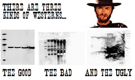
Need some help to tame that wild western blot? Here are some tips and tricks to help you along the way –

  • Always check ladder migration pattern based on specific gel and electrophoresis conditions, as these factors can shift the apparent molecular weights from the supposed “standard” ladder image given out by the company.
  • When testing a new antibody, leave the blot intact, opting to strip it and perform a control protein blot after probing for your target protein. Cutting the blot and using different pieces for your target and control protein on a first try may obscure alternative target protein isoforms or off-target background staining.
  • High background? Try a more stringent blocking condition than just BSA or milk by adding goat serum or fish gelatin to your solution. Blocking overnight also can help clean up your blots.
  • Is you gel “smiling” or “frowning”? This usually happens when your sample buffer has too much salt.
  • Make sure your PVDF membrane is pre-activated with methanol for 20 minutes before making gel sandwich. It’s also a good idea to mark which side of the membrane is facing the gel with a sharpie, on a top corner.
  • It’s always a good idea to do a Ponceau stain on your membrane after transfer to make sure your transfer went all right. Alternatively, you can also stain your gel with Coomassie blue.
  • It is possible to over-transfer, especially for low MW proteins (<10 kDa) – optimize transfer time or reduce voltage. On the other hand, high MW proteins will take longer time to transfer.
  • Have a dirty secondary? Consider adding a wee-bit of Tween-20 to your washes (0.01-0.5%).
  • When loading your samples, press on the pipet just enough to get any possible air bubbles out and run the tip through the running buffer in the tank before putting it in the well. And make sure your sample was denatured prior to running.
  • If power supply reading shows 0 when switched on, make sure your power cables are properly connected to the power supply. If that doesn’t work, check for broken electrodes or blown fuses. Lastly, try with a higher limit power supply.
  • As always, make sure you write down the protocol before-hand and check through every step when performing. This will help you track your steps back to see at which step things could have gone wrong.


Source - http://prooffreaderplus.blogspot.com/2014/05/one-of-my-favourite-memories-good-bad.html
Source – http://prooffreaderplus.blogspot.com/2014/05/one-of-my-favourite-memories-good-bad.html