Optical Tweezers
Optical Tweezer at crosshairs moving microsphere in alginate gel. The softer the gel, the more the microsphere moves
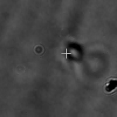
Optical Tweezer at crosshairs moving microsphere in alginate gel. The softer the gel, the more the microsphere moves
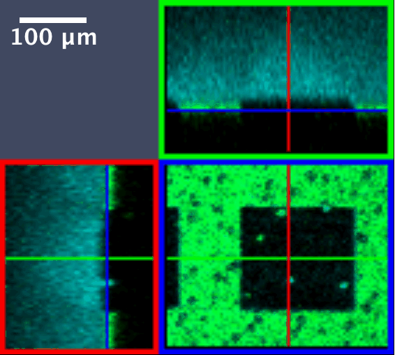
Ultrafast beam in nonlinear optical microscope used to write patterns in azobenzene modified silk

Novel material combining advantages of deep eutectic solvent systems and polyol-citric acid polycondensates. Here used as a light guide.
My research is based on the interactions of light and matter, and its use for biomedical applications. These interactions range from the use of light to pattern biomaterials to optical tweezers, in which the electric fields of tightly focussed laser light can apply appreciable forces to microscopic materials: cells in culture or microspheres used as probes of the stiffness and viscoelastic response of extracellular matrix. This enable us to non-invasively probe the effects of mechanical stimuli.
During my research leave at the Optics Laboratory, EPFL Lausanne I researched the use of deep learning to image through bio-friendly fibers such as gelatin supported deep eutectic solvents (DES) and silk. The main outcome was the development of DES assisted, bio-friendly polyesters based on polyols such as 1,8-octanediol and citric acid. My current research concentrates on optical beam propagation in nonlinear optical materials.
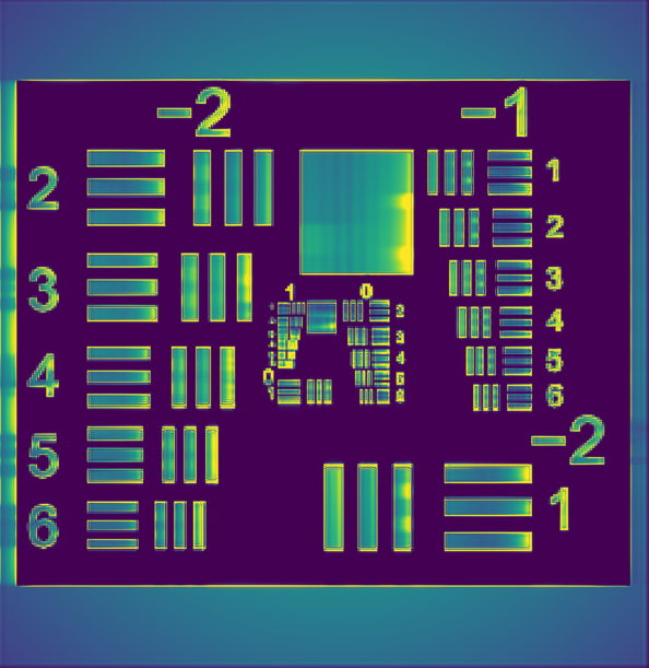
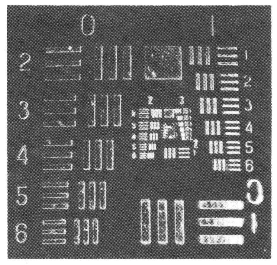
A lot of the theoretical models of beam amplification in nonlinear optical materials had been carried out in one transverse spatial dimension because of computing limitations in the 90s. Modern computers and GPUs have enabled full three dimensional time dependent models of these processes. My lab has recently completed open source software for these computations. Typical outputs are shown below: Left to right: photorefractive optical soliton, computed image amplification showing edge enhancement distortions due to saturation, corresponding experimentally observed output. Read more by following the research tab above.