Photorefractive beam coupling software
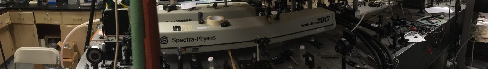
In the early 1990’s when compute power was limited I wrote a software package to model optical interactions in photorefractive materials such as barium titanate. With the subsequent rapid improvement in the state of the art in computing software and hardware I have reworked the software into its modern equivalent: the photorefractive beam coupler. This software can be used to model a variety of photorefractive effects and devices. Photorefractive materials are real time holographic recording materials that can be used to amplify image bearing beams, correct for optical distortions, and support spatial solitons. Several types of ferroelectric crystals such as barium titanate, potassium niobate and strontium barium niobate have Curie transitions near room temperature and are especially susceptible to optically induced changes in refractive index. These changes affect the propagation of laser beams as they pass through such a crystal. For example when a single beam passes through a half centimeter cube of barium titanate, some of it is scattered from microscopic imperfections in the crystal. Interference between this scattered light and the main beam is automatically recorded as a hologram This hologram diffracts light from the main beam into the scattered light, thereby amplifying it in an effect commonly known as fanning. If you place a white screen you will see a broad fan of bight scattered light. The figure below shows an actual fanning pattern on the left and a computed version of the corresponding beam in the crystal on the right.


Optical Tweezers
In 1970, Arthur Ashkin reported that spatial variations in beams of light could apply forces to microscopic particles. Since that discovery, this effect, now known as optical tweezers has enabled the growth of an active research community developing the theory and application of optical micromanipulation, especially in the context of biology and biomedical engineering where the objects of interest: cells, bacteria, and even DNA are at a size scale suitable for non-invasive probing and manipulation by suitably prepared beams of light. The light that is used is often in the form of tightly focussed laser beams or beams with complex structure generated by spatial light modulators. In 1996 Professor Cronin-Golomb spent a sabbatical at Stanford University in Steve Chu’s lab where he first gained experience with optical tweezers and noted its relationship to photorefractive nonlinear optics, his first area of research.
At Tufts, the lab’s main line of research has been in the use of optical tweezers for non-invasive measurement of the viscoelastic and rheological properties of fluids and biomaterials, most recently in a collaboration with Matt Panzer and Sameer Sonkusale on the mechanical properties of a biofriendly low vapor pressure gel. An earlier collaboration was with Irene Georgakoudi and Ana Soto for spatially resolved measurements of the stiffness of the extracellular matrix in three dimensional cultures of mammary gland tissue. This is for the purpose of gaining an understanding of the mechanical cues thought to play a substantial role in determining cell differentiation and mammary gland development in the context of cancer research. The approach involves seeding the cell culture with polystyrene or silica microspheres for use as probe particles. The response of these embedded particles to forces applied by optical tweezers is used to measure local mechanical properties such as Young’s modulus.
Information about the setup and associated software can be found in this GitHub repository.
Video walkthrough of optical layout
Our fully equipped optics lab includes an inverted microscope optical tweezers apparatus with a Labview enabled computer interface which provides opportunities for research in automatic particle tracking and measurement using image analysis and closed loop tracking via galvanometer and piezoelectric beam steering. Measurements are made via phase sensitive detection of forward and backscattered light from micron sized probe particles embedded in the matrix. We have also been using one of Irene Georgakoudi’s two-photon scanning microscopes to use the signal from fluorescently tagged probe particles, while simultaneously imaging the collagen matrix via second harmonic generation.
At the same time, we are developing theoretical background for the measurements by using finite element techniques to model the reponse of microspheres to optical forces in materials with known rheological properties.
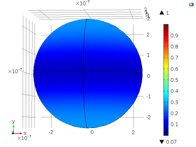
Tweezing a bead in gel
Click on the image to see an animated model of a 0.25 micron polystryrene sphere forced by a 25mW near infrared laser beam in a soft gel.
Nonlinear Optics in Biomaterials
There is a long history of the use of the photoisomerizable molecule azobenzene to photosensitize polymers for optical recording and optical holography. In 2008 Amanda Murphy, Peter St. John, and David Kaplan: members of the silk group in our department showed that the tyrosine amino acids in silk fibroin could be functionalized via diazonium coupling to form azobenzene side groups. In 2012 we showed that thin films of silk functionalized in this way behaved in the same way as previously studied azo-polymers. Azosilk shows optically induced birefringence and posesses the ability to act as a holographic recording medium, complete with the surface relief gratings commonly associated with holographic recording in other azopolymers. We were also able to show that a silk-elastin like protein (SELP) could be functionalized with retinal via linkage to the amino acid lysine in the SELP backbone. Retinal, like azobenzene, is photoisomerizable and displays many of the same effects as azobenzene, but is more biocompatible: it forms the basis of the visual system. In 2013-2014 Professor Cronin-Golomb spent a sabbatical year at McGill University in the Chemistry Department with Professor Christopher Barrett, an expert in azobenzene chemistry and optics. There, a collaboration with McGill’s Advanced BioImaging Facility resulted in the demonstration of the use of a two-photon microscope to make lithographically patterned modifications to azosilk film gels. These modifications include fluorescence patterning and the production of hollow microfuidic chambers suitable for developing microfluidic devices and surface patterning to guide cell growth.
Photoliquefiable materials for drug delivery
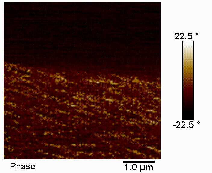
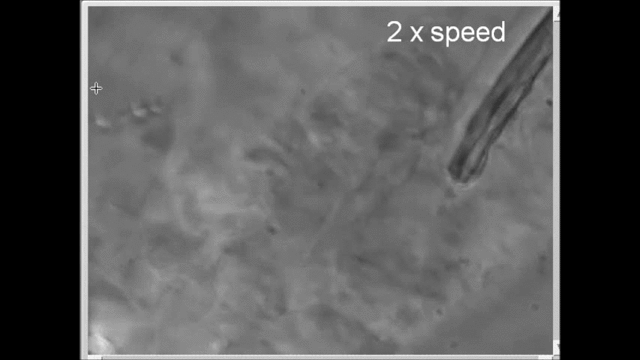
Photoliquefiable ionic crystals are solids that become liquid when illuminated with ultraviolet light and solidify again via thermal relaxation or illumination with blue light. These materials first gained attention for use as energy storage materials. Lorenz Meinel’s group at the University of Würtzburg has been considering a quite different application optically tunable transdermal delivery of pharmaceuticals. We have been collaborating with that group to learn more about the properties of these materials. One problem with using ultraviolet activation is that the optical penetration depth is limited. By using two photon activation, we could simultaneously improve the penetration depth and target delivery more precisely to a given spatial location. Below, we show results from our lab using atomic force microscopy, as well demonstrations of phase transitions induced by two-photon excitation.
Rheological properties of gelatin supported deep eutectic solvents
In a collaboration with Matt Panzer in the Department of Chemical and Biological Engineering we have begun to investigate the mechanical properties of transparent gel materials using gelatin in a deep eutectic like host material comprising the bio-friendly materials choline chloride (used in chicken feed) and propanediol (used in cosmetics). Eutectics are mixtures of materials whose melting points are lower than that any of the ingredients. While the original motivation for studying these materials was to use them as electrolytes in supercapacitors, the observation that they are transparent soft gels prompts the question of whether they can be used in applications requiring optical biomaterials. The vapor pressure of this material is low enough that it does not readily dry out in ambient air. The aim of this project is to measure its micro-rheological properties. Because the material includes gelatin protein implies that it may be structurally inhomogeneous, so micro-rheological measurements may useful.
DES assisted polycondensates for bio-optics
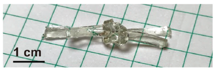
Current progress in imaging through multimode fibers suggests revisiting endoscopic imaging and controlled light delivery in biocompatible and biodegradable fibers. Previous research on waveguiding in biofibers has included biofriendly biodegradable materials such as silkworm silk and crosslinked polycondensates such as poly(octanediol-co-citrate) (POC). At the same time, ecofriendly deep eutectic solvent (DES) materials are being developed for energy storage applications. By themselves, they are generally in liquid form or are brittle solids and so do not form flexible soft materials amenable to formation into optical fibers with mechanical properties compatible with human tissue. Panzer et al have shown that gelatin added to choline chloride-based DES can provide support enabling the formation of gels suitable for making flexible, wearable conductive sensors. One of the aims of this project was to investigate the possibility of using DES and OCT/POC polycondensate materials to make optical waveguides and fibers. Both of these material classes have some problems to be overcome. The former degrades rapidly in aqueous solutions thus limiting biomedical application, and the latter requires processing temperatures well above 100oC. The production of POC involves the reaction of 1,8-octanediol with citric acid at 140oC. But we can combine the best of both types of material by first adding choline chloride to 1,8-octanediol to form a DES to lower the melting point, then carrying out a reaction with citric acid to form a DES POC at 90oC. Below, we report the results of our research on DES POC type materials and a short parallel effort using silkworm silk fibroin which has a good track record in biomedical devices, tissue engineering and biocompatible optics and sensors.