Main Motivations
The TimkoLab focuses on two-way communication with electrically-active cells, monitoring tissue function for long periods of time. Key focuses of research include the usage of biomaterials to develop seamless devices for interfacing and recording biological processes.
Project 1: Heart-On-A-Chip
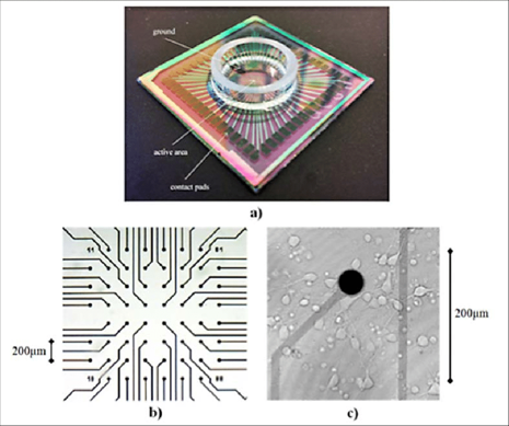
This project is targeted at monitoring and modeling of cardiac disease through bioelectric devices – more specifically, action potential modeling of cardiac tissue through multielectrode array construction with HL-1 Cells
- Xie, C., Lin, Z., Hanson, L. et al. Intracellular recording of action potentials by nanopillar electroporation. Nature Nanotech 7, 185–190 (2012).
- Contacts: Haitao Liu, PhD | Rotimi Bolonduro | Akshita Rao (Class of 2021)
One major area of investigation using these modelling technologies focuses on hypoxic stress and changes in HL-1 (cardiomyocyte) cell behavior:
- Investigation of hypoxic stress and changes in HL-1 cell behavior
- Hypoxia-inducible factor-1 alpha (HIF-1α) facilitates metabolic adaptation by translocating into nucleus, binding HIF-1β, and affecting gene upregulation.
- However, HIF-1α rapidly degrades upon exposure to normal oxygen conditions.
- The development of the above extracellular multielectrode array allows long-term monitoring without compromising cell integrity or introducing confounding variables.
By multiplexing the readouts to larger cardiomyocyte monolayers, research revealed the generation of an action potential “wave” that drove synchronized contraction. This wave was disrupted in an hypoxic environment:

and future research showed that, on a single-cell basis, the shape and behavior of action potentials changed depending on outside oxygen concentration.
Project 2: Silk Fibroin for Bioelectrical Applications
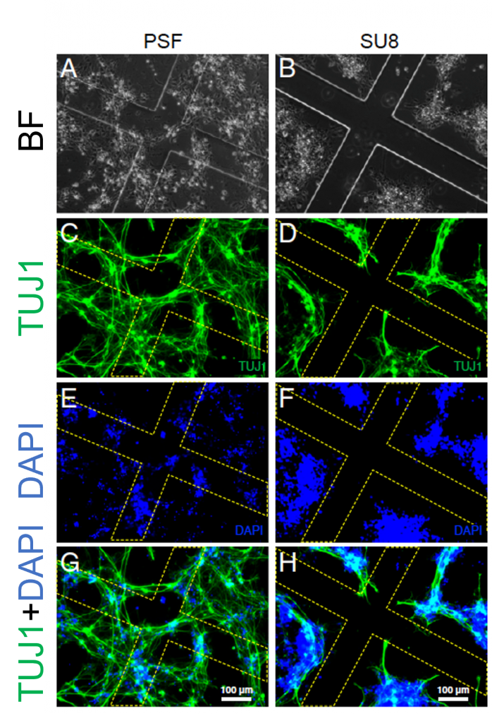
The current gold standard of bioelectronic foundation lies in a material called SU8, which is a photo-crosslinkable polymer. However, this material can suffer from biomechanical mismatches in implanted devices. Thus, the TimkoLab turned to silk!
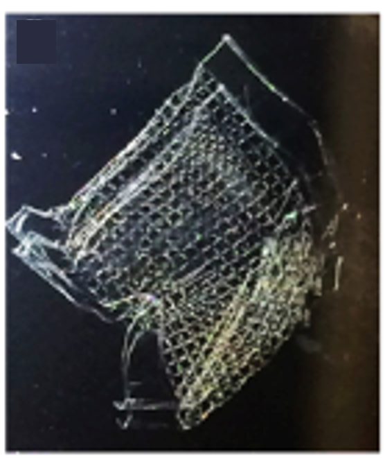
Methylacrylation of silk imparts photosensitivity, which creates silk fibroin that is crosslinkable (allowing light-based hardening of the solution) – known as photocrosslinkable silk fibroin (PSF):
- PSF is compatible with conventional photolithography (3D printing, etc.)
- PSF also allows for structures with tunable thickness, transparent, freestanding, high precision (1 micron structure resolution)
- PSF also acts as an insulator, allowing it to act as a bioelectric foundation for devices
- Contact — Jie Ju, PhD
Further experiments with PSF revealed that although cells do not like to attach to it normally, this is due to the “wettability” of the material. By adding polylysine groups to the construction of PSF, TimkoLab figured out a way to increase the wettability of PSF in certain areas, incentivizing adhesion to printed areas:
To learn more about these projects or ways to get involved, contact Brian Timko:
https://engineering.tufts.edu/bme/people/faculty/brian-timko