Final Poster Presentation
Reply

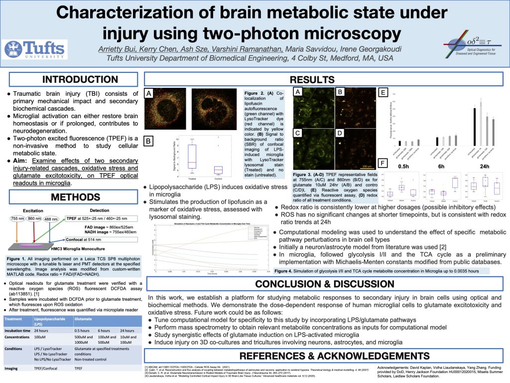
Introduction
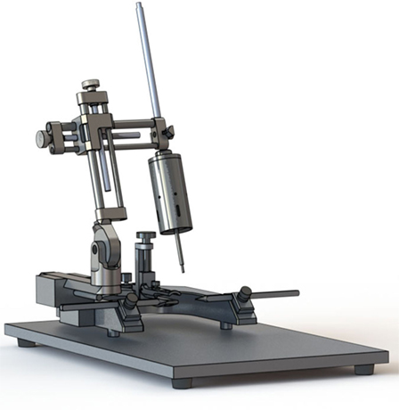
Traumatic brain injury (TBI) is a leading cause of death and disability worldwide. However, TBI remain difficult to identify and treat in the clinic due to a lack of known biomarkers that can be used as diagnostic and therapeutic targets. To this end, 3D engineered brain tissues are seeded with human induced neuronal stem cells (hINSCs), and they are assessed using two-photon excited fluorescence (TPEF). With the results from imaging, we aim to correlate such optical measurements with biochemical and metabolomic assays in the context of two major aspects of TBI, glutamate excitotoxicity and oxidative stress. This work will ultimately be used to develop a metabolic model that will use optical measurements to identify biomarkers that are implicated in TBI-associated pathways.
Aim: Examine effects of two secondary injury-related cascades, oxidative stress and glutamate excitotoxicity, on TPEF optical readouts in microglia.
In order to achieve this aim, we treated engineered microglia with lipopolysaccharide and LPS. We quantified fluorescence and imaged the tissue with TPEF after. Our experiments lead us to the following results:
● Lipopolysaccharide (LPS) induces oxidative stress in microglia and stimulates the production of lipofuscin as a marker of oxidative stress, assessed with lysosomal staining.
● Redox ratio is consistently lower at higher dosages
● The computational model can help us understand glycolysis I/II and the TCA cycle in microglia but will need further work to fully integrate experimental results
In our work, we establish a platform for studying metabolic responses to secondary injury in brain cells using optical and biochemical methods. We demonstrate the dose-dependent response of human microglial cells to glutamate excitotoxicity and
oxidative stress.
Future work could be as follows:
● Tune computational model for specificity to this study by incorporating LPS/glutamate pathways
● Perform mass spectrometry to obtain relevant metabolite concentrations as inputs for computational model
● Study synergistic effects of glutamate induction on LPS-activated microglia
● Induce injury on 3D co-cultures and tricultures involving neurons, astrocytes, and microglia