General Opioid Signaling
Pain signal inhibition: Our body produces natural (endogenous) opioids, which comprise endorphins, dynorphins, and enkephalins (4). On a molecular level, both these and synthetic opioids inhibit pain signals by interfering with intracellular signaling and preventing ascending pain-causing neurons from sending action potentials. In this section we describe how opioid binding inhibit the transmission of neuronal signals in pain-activated neurons though hyperpolarizing the cell membrane, inhibiting adenylyl cyclase and preventing neurotransmitter release.
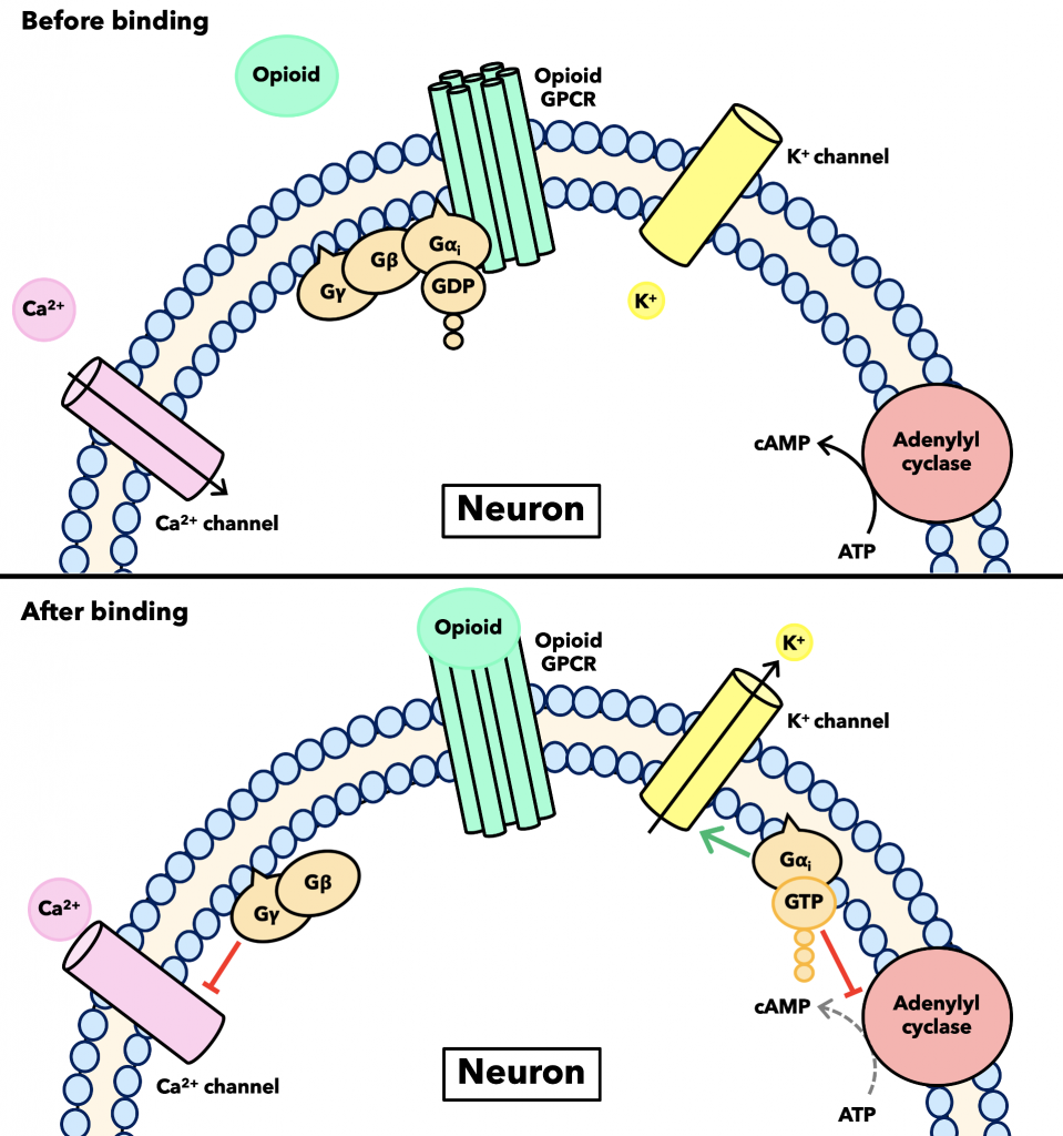
Hyperpolarization & adenylyl cyclase inhibition: This process begins when opioids bind to either µ, κ, δ or opioid receptor-like 1 opioid receptors, which are all G-protein coupled receptors (GPCRs). This means that once the opioid ligand binds, the Gαi subunit undergoes a conformational change that decreases its affinity for GDP and increases it for GTP, causing the former to be switched out for the latter (4). This exchange yields another conformational change that causes the dissociation of the coupled G-protein into its Gαi and Gβγ subunits, which break off from the receptor. The Gαi subunit goes on to inhibit the production of cAMP, a secondary messenger that has been shown to be important in maintaining peripheral hyperalgesia (4), by binding to and inhibiting the ATP to cAMP-converting enzyme adenylyl cyclase. HThe Gαi subunit also opens the Kir3 potassium ion channel, allowing positively charged potassium ions to flow down their concentration gradient out of the cell (4). This hyperpolarizes the membrane, inhibiting the generation and propagation of an action potential since action potentials are triggered only when the membrane potential is sufficiently depolarized.

Neurotransmitter release inhibition: The inhibitory effect is compounded though opioid binding preventing the release of neurotransmitters by affected neurons. This happens though the the Gβγ subunit binding to and closing calcium channels, preventing calcium ions from entering the cell. Neurons communicate with one another via neurotransmitters being released from one neuron (the presynaptic neuron) and binding to receptors on another (the postsynaptic neuron) (13). Neurotransmitters are initially contained within vesicles, and their release occurs when those vesicles fuse with the presynaptic membrane. Calcium ions promote this fusion, so blocking their entry prevents the chemical transmission of neuronal signals across synapses as well as hyperpolarizes the cell membrane (Figure 8) (4). See an example of how this inhibition impacts GABA signaling in the µ receptors section. Opioid binding has several other downstream effects, but what is discussed here are the principal modes of neuronal inhibition.
November 8, 2019 at 5:27 pm
I think the paragraphs would make more sense to me personally if they were broken up a little bit more into smaller ones. The amount of different proteins that you have to talk about get all jumbled in my head and its hard to keep them straight. The image does a good job of helping me picture it, but I think breaking it up would make it clearer. The last sentence you have describing the mu receptors is really nice to tie it all in and bring you to the next page.
December 14, 2019 at 9:20 pm
I split up the introduction into pain inhibition and the specific description of the GCPR figure into two separate sections separated the figure. Thanks for the advice!
November 10, 2019 at 4:31 pm
Is there a citation for the first figure? I agree with Paige- putting the figure in the middle of the very technical sequence of protein and subunit binding would help to smooth out the reading. As well, the first section could use a comparison to normal neuronal function- in the second section, there is a good example of normal vs inhibited calcium ion activity, and I think as an audience who isn’t super familiar with the pathway it would be helpful to highlight which things change with opioids bound. It seems that the main inhibition of signals is constant hyperpolarization, in which case that should be stressed as it’s not super clear.
December 14, 2019 at 9:20 pm
I actually made the first picture so it does not need a citation 🙂 Also in the new separated introduction section I added a part that states clearly the three inhibitory affects opioid binding cause. Finally I added new headings to each of the sections in bold to describe which inhibitory affect is being examined here. Hope the three modes of inhibition discussed are more clear!
November 10, 2019 at 5:50 pm
Very information dense again, but the pictures are very informative.
December 14, 2019 at 9:20 pm
Molecular mechanism in general can be really information dense since they often involve many simultaneous processes but I’m glad to hear that the diagrams help you digest the information.
November 10, 2019 at 7:31 pm
All of the information is really interesting but I think that most importantly the paragraphs flow extremely well. The first section was hard to understand but the image was helpful in seeing what was happening. I found that there the image and paragraph worked together to increase understanding. The second image and paragraph were both interesting but I think that I found it harder to understand the second image from the paragraph. I just didn’t think that the image and paragraph built off of each other as the first paragraph and image did.
December 14, 2019 at 9:21 pm
The second image is just used to show on a cellular scale how the binding of opioids at one section can alter the release of neurotransmitters at a completely separate section. I think it is definitely necessary to keep in since it provides this larger perspective and helps more visual learners picture how neurotransmitter release is inhibited.